Artículo
Fluorescent immunosensor using AP-SNs and QDs for quantitation of IgG anti-Toxocara canis
Medawar Aguilar, Victoria ; Messina, Germán Alejandro
 ; Fernández Baldo, Martín Alejandro
 ; Raba, Julio
 ; Pereira, Sirley Vanesa
 ; Messina, Germán Alejandro
 ; Fernández Baldo, Martín Alejandro
 ; Raba, Julio
 ; Pereira, Sirley Vanesa
Fecha de publicación:
01/2017
Editorial:
Elsevier Science
Revista:
Microchemical Journal
ISSN:
0026-265X
Idioma:
Inglés
Tipo de recurso:
Artículo publicado
Clasificación temática:
Resumen
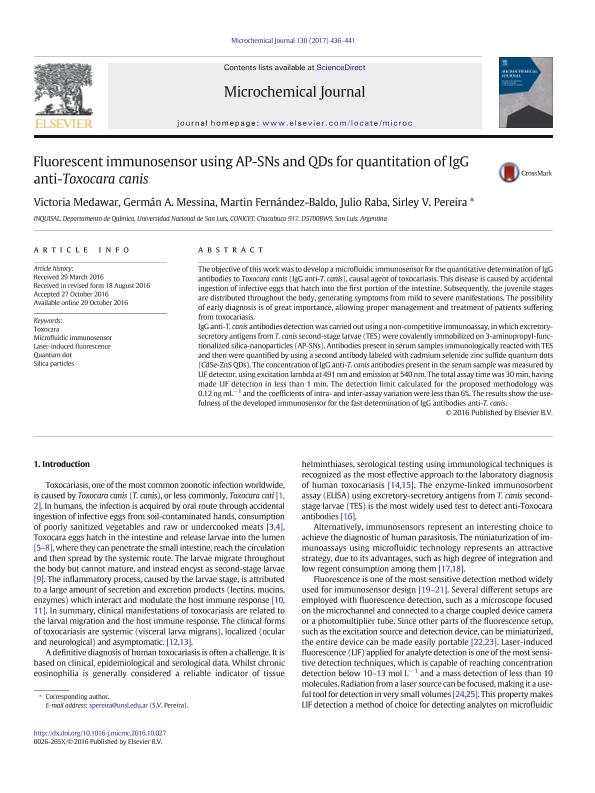
The objective of this work was to develop a microfluidic immunosensor for the quantitative determination of IgG antibodies to Toxocara canis (IgG anti-T.canis), causal agent of toxocariasis. This disease is caused by accidental ingestion of infective eggs that hatch into the first portion of the intestine. Subsequently, the juvenile stages are distributed throughout the body, generating symptoms from mild to severe manifestations. The possibility of early diagnosis is of great importance, allowing proper management and treatment of patients suffering from toxocariasis. IgG anti-T.canis antibodies detection was carried out using a non-competitive immunoassay, in which excretory-secretory antigens from T. canis second-stage larvae (TES) were covalently immobilized on 3-aminopropyl-functionalized silica-nanoparticles (AP-SNs). Antibodies present in serum samples immunologically reacted with TES and then were quantified by using a second antibody labeled with cadmium selenide zinc sulfide quantum dots (CdSe-ZnS QDs). The concentration of IgG anti-T. canis antibodies present in the serum sample was measured by LIF detector, using excitation lambda at 491 nm and emission at 540 nm. The total assay time was 30 minutes, having made LIF detection in less than 1 minute. The detection limit calculated for the proposed methodology was 0.12 ng mL-1 and the coefficients of intra- and inter-assay variation were less than 6%. The results show the usefulness of the developed immunosensor for the fast determination of IgG antibodies anti T. canis.
Archivos asociados
Licencia
Identificadores
Colecciones
Articulos(INQUISAL)
Articulos de INST. DE QUIMICA DE SAN LUIS
Articulos de INST. DE QUIMICA DE SAN LUIS
Citación
Medawar Aguilar, Victoria; Messina, Germán Alejandro; Fernández Baldo, Martín Alejandro; Raba, Julio; Pereira, Sirley Vanesa; Fluorescent immunosensor using AP-SNs and QDs for quantitation of IgG anti-Toxocara canis; Elsevier Science; Microchemical Journal; 130; 1-2017; 436-441
Compartir
Altmétricas


