Artículo
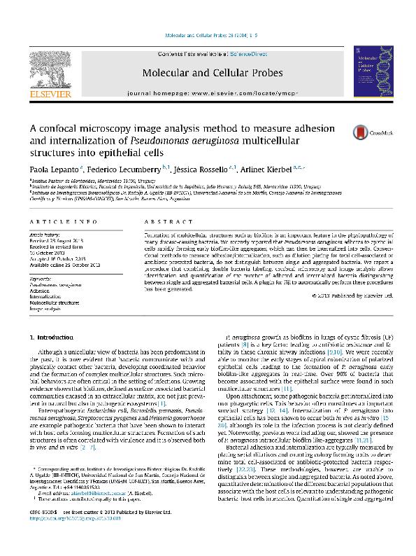
A confocal microscopy image analysis method to measure adhesion and internalization of Pseudomonas aeruginosa multicellular structures into epithelial cells
Fecha de publicación:
02/2014
Editorial:
Academic Press Ltd-elsevier Science Ltd
Revista:
Molecular And Cellular Probes
ISSN:
0890-8508
Idioma:
Inglés
Tipo de recurso:
Artículo publicado
Clasificación temática:
Resumen
Formation of multicellular structures such as biofilms is an important feature in the physiopathology of many disease-causing bacteria. We recently reported that Pseudomonas aeruginosa adheres to epithelial cells rapidly forming early biofilm-like aggregates, which can then be internalized into cells. Conventional methods to measure adhesion/internalization, such as dilution plating for total cell-associated or antibiotic protected bacteria, do not distinguish between single and aggregated bacteria. We report a procedure that combining double bacteria labeling, confocal microscopy and image analysis allows identification and quantification of the number of adhered and internalized bacteria distinguishing between single and aggregated bacterial cells. A plugin for Fiji to automatically perform these procedures has been generated.
Archivos asociados
Licencia
Identificadores
Colecciones
Articulos(IIB-INTECH)
Articulos de INST.DE INVEST.BIOTECNOLOGICAS - INSTITUTO TECNOLOGICO CHASCOMUS
Articulos de INST.DE INVEST.BIOTECNOLOGICAS - INSTITUTO TECNOLOGICO CHASCOMUS
Citación
Kierbel, Arlinet Verónica; Rossello, Jéssica; Lecumberry, Federico; Lepanto, Paola; A confocal microscopy image analysis method to measure adhesion and internalization of Pseudomonas aeruginosa multicellular structures into epithelial cells; Academic Press Ltd-elsevier Science Ltd; Molecular And Cellular Probes; 28; 1; 2-2014; 1-5
Compartir
Altmétricas


