Artículo
Impact of manganese on primary hippocampal neurons from rodents
Daoust, Alexia; Saoudi, Yasmina; Brocard, Jacques; Collomb, Nora; Batandier, Cecile; Bisbal, Mariano ; Salomé, Murielle; Andrieux, Annie; Bohic, Sylvain; Barbier , Emmanuel
 ; Salomé, Murielle; Andrieux, Annie; Bohic, Sylvain; Barbier , Emmanuel
Fecha de publicación:
05/2014
Editorial:
Wiley-liss, Div John Wiley & Sons Inc
Revista:
Hippocampus
ISSN:
1050-9631
Idioma:
Inglés
Tipo de recurso:
Artículo publicado
Clasificación temática:
Resumen
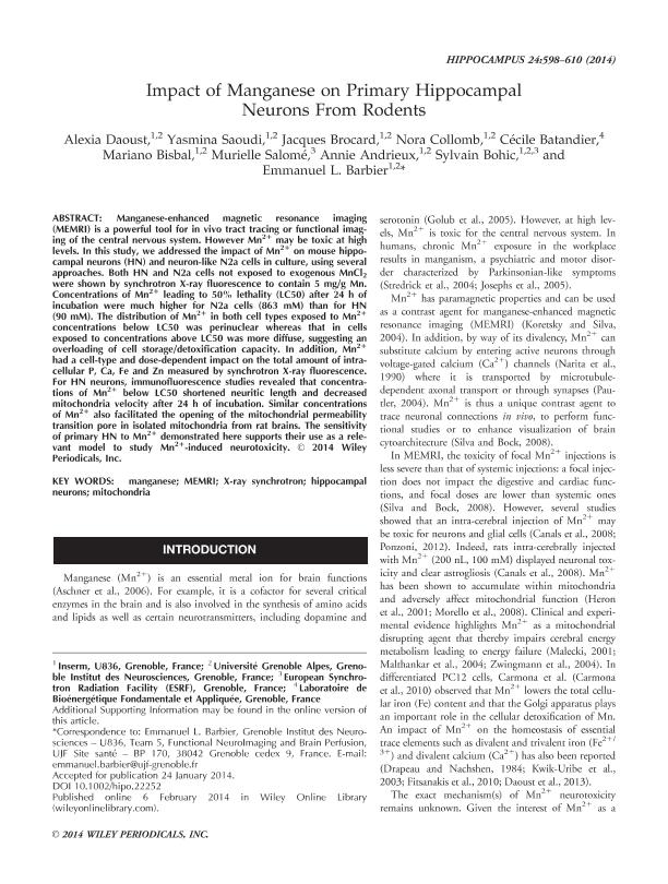
Manganese-enhanced magnetic resonance imaging (MEMRI) is a powerful tool for in vivo tract tracing or functional imaging of the central nervous system. However Mn2+ may be toxic at high levels. In this study, we addressed the impact of Mn2+ on mouse hippocampal neurons (HN) and neuron-like N2a cells in culture, using several approaches. Both HN and N2a cells not exposed to exogenous MnCl2 were shown by synchrotron X-ray fluorescence to contain 5 mg/g Mn. Concentrations of Mn2+ leading to 50% lethality (LC50) after 24 h of incubation were much higher for N2a cells (863 mM) than for HN (90 mM). The distribution of Mn2+ in both cell types exposed to Mn2+ concentrations below LC50 was perinuclear whereas that in cells exposed to concentrations above LC50 was more diffuse, suggesting an overloading of cell storage/detoxification capacity. In addition, Mn2+ had a cell-type and dose-dependent impact on the total amount of intracellular P, Ca, Fe and Zn measured by synchrotron X-ray fluorescence. For HN neurons, immunofluorescence studies revealed that concentrations of Mn2+ below LC50 shortened neuritic length and decreased mitochondria velocity after 24 h of incubation. Similar concentrations of Mn2+ also facilitated the opening of the mitochondrial permeability transition pore in isolated mitochondria from rat brains. The sensitivity of primary HN to Mn2+ demonstrated here supports their use as a relevant model to study Mn2+-induced neurotoxicity.
Palabras clave:
Manganese
,
Memri
,
X-Ray Synchrotron
,
Hippocampalneurons
,
Mitochondria
Archivos asociados
Licencia
Identificadores
Colecciones
Articulos(INIMEC - CONICET)
Articulos de INSTITUTO DE INV. MEDICAS MERCEDES Y MARTIN FERREYRA
Articulos de INSTITUTO DE INV. MEDICAS MERCEDES Y MARTIN FERREYRA
Citación
Barbier , Emmanuel; Andrieux, Annie; Salomé, Murielle; Bisbal, Mariano; Batandier, Cecile; Collomb, Nora; et al.; Impact of manganese on primary hippocampal neurons from rodents; Wiley-liss, Div John Wiley & Sons Inc; Hippocampus; 24; 5; 5-2014; 598-610
Compartir
Altmétricas


