Mostrar el registro sencillo del ítem
dc.contributor.author
Diessler, Mónica Elizabeth

dc.contributor.author
Hernández, Rocío

dc.contributor.author
Gomez Castro, María Gimena

dc.contributor.author
Barbeito, Claudio Gustavo

dc.date.available
2023-12-28T15:58:53Z
dc.date.issued
2023-03
dc.identifier.citation
Diessler, Mónica Elizabeth; Hernández, Rocío; Gomez Castro, María Gimena; Barbeito, Claudio Gustavo; Decidual cells and decidualization in the carnivoran endotheliochorial placenta; Frontiers Media S. A.; Frontiers in Cell and Developmental Biology; 11; 3-2023; 1-16
dc.identifier.issn
2296-634X
dc.identifier.uri
http://hdl.handle.net/11336/221832
dc.description.abstract
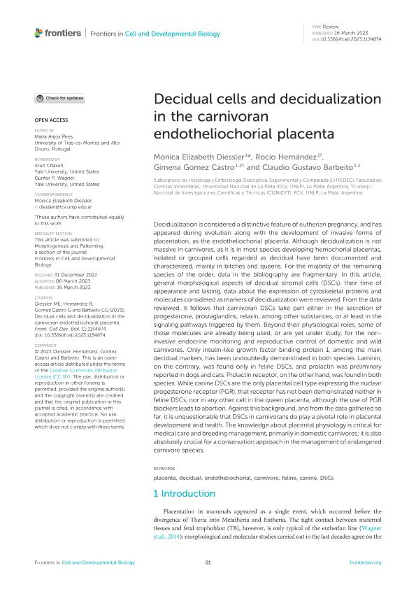
Decidualization is considered a distinctive feature of eutherian pregnancy, and has appeared during evolution along with the development of invasive forms of placentation, as the endotheliochorial placenta. Although decidualization is not massive in carnivores, as it is in most species developing hemochorial placentas, isolated or grouped cells regarded as decidual have been documented and characterized, mainly in bitches and queens. For the majority of the remaining species of the order, data in the bibliography are fragmentary. In this article, general morphological aspects of decidual stromal cells (DSCs), their time of appearance and lasting, data about the expression of cytoskeletal proteins and molecules considered as markers of decidualization were reviewed. From the data reviewed, it follows that carnivoran DSCs take part either in the secretion of progesterone, prostaglandins, relaxin, among other substances, or at least in the signaling pathways triggered by them. Beyond their physiological roles, some of those molecules are already being used, or are yet under study, for the non-invasive endocrine monitoring and reproductive control of domestic and wild carnivores. Only insulin-like growth factor binding protein 1, among the main decidual markers, has been undoubtedly demonstrated in both species. Laminin, on the contrary, was found only in feline DSCs, and prolactin was preliminary reported in dogs and cats. Prolactin receptor, on the other hand, was found in both species. While canine DSCs are the only placental cell type expressing the nuclear progesterone receptor (PGR), that receptor has not been demonstrated neither in feline DSCs, nor in any other cell in the queen placenta, although the use of PGR blockers leads to abortion. Against this background, and from the data gathered so far, it is unquestionable that DSCs in carnivorans do play a pivotal role in placental development and health. The knowledge about placental physiology is critical for medical care and breeding management, primarily in domestic carnivores; it is also absolutely crucial for a conservation approach in the management of endangered carnivore species.
dc.format
application/pdf
dc.language.iso
eng
dc.publisher
Frontiers Media S. A.
dc.rights
info:eu-repo/semantics/openAccess
dc.rights.uri
https://creativecommons.org/licenses/by/2.5/ar/
dc.subject
CANINE
dc.subject
CARNIVORE
dc.subject
DECIDUAL
dc.subject
DSCS
dc.subject
ENDOTHELIOCHORIAL
dc.subject
FELINE
dc.subject
PLACENTA
dc.subject.classification
Biología del Desarrollo

dc.subject.classification
Ciencias Biológicas

dc.subject.classification
CIENCIAS NATURALES Y EXACTAS

dc.subject.classification
Biología Reproductiva

dc.subject.classification
Ciencias Biológicas

dc.subject.classification
CIENCIAS NATURALES Y EXACTAS

dc.title
Decidual cells and decidualization in the carnivoran endotheliochorial placenta
dc.type
info:eu-repo/semantics/article
dc.type
info:ar-repo/semantics/artículo
dc.type
info:eu-repo/semantics/publishedVersion
dc.date.updated
2023-12-27T17:39:55Z
dc.journal.volume
11
dc.journal.pagination
1-16
dc.journal.pais
Suiza

dc.description.fil
Fil: Diessler, Mónica Elizabeth. Universidad Nacional de la Plata. Facultad de Ciencias Veterinarias. Laboratorio de Histología y Embriología Descriptiva, Experimental y Comparada (LHYEDEC);
dc.description.fil
Fil: Hernández, Rocío. Universidad Nacional de la Plata. Facultad de Ciencias Veterinarias. Laboratorio de Histología y Embriología Descriptiva, Experimental y Comparada (LHYEDEC); . Consejo Nacional de Investigaciones Científicas y Técnicas. Centro Científico Tecnológico Conicet - La Plata; Argentina
dc.description.fil
Fil: Gomez Castro, María Gimena. Universidad Nacional de la Plata. Facultad de Ciencias Veterinarias. Laboratorio de Histología y Embriología Descriptiva, Experimental y Comparada (LHYEDEC); . Consejo Nacional de Investigaciones Científicas y Técnicas. Centro Científico Tecnológico Conicet - La Plata; Argentina
dc.description.fil
Fil: Barbeito, Claudio Gustavo. Universidad Nacional de la Plata. Facultad de Ciencias Veterinarias. Laboratorio de Histología y Embriología Descriptiva, Experimental y Comparada (LHYEDEC); . Consejo Nacional de Investigaciones Científicas y Técnicas. Centro Científico Tecnológico Conicet - La Plata; Argentina
dc.journal.title
Frontiers in Cell and Developmental Biology
dc.relation.alternativeid
info:eu-repo/semantics/altIdentifier/url/https://www.frontiersin.org/articles/10.3389/fcell.2023.1134874/full
dc.relation.alternativeid
info:eu-repo/semantics/altIdentifier/doi/http://dx.doi.org/10.3389/fcell.2023.1134874
Archivos asociados
