Artículo
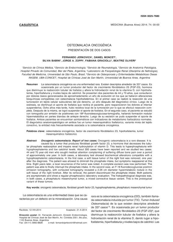
La osteomalacia oncogénica es una enfermedad rara. Existen descriptos alrededor de 337 casos. Es ocasionada por un tumor productor del factor de crecimiento fibroblástico 23 (FGF-23), hormona que disminuye la reabsorción tubular de fosfatos y altera la hidroxilación renal de la vitamina D, con hipofosfatemia, hiperfosfaturia y niveles bajos de calcitriol. Se presentan dos pacientes de 44 y 70 años, que consultaron por dolores óseos generalizados de aproximadamente un año de evolución en los que se hallaron alteraciones bioquímicas compatibles con osteomalacia hipofosfatémica. En el primer caso se realizó la resección de una tumoración en tejido celular subcutáneo del pie derecho, un año después del diagnóstico clínico. Luego de la exéresis, se disminuyó el aporte de fosfatos que recibía el paciente, pero reaparecieron los dolores al intentar suspenderlos. Ocho años más tarde, hubo recidiva local de la tumoración por lo que se efectuó resección completa. Después de la misma, se logró suspender el aporte de fosfatos. En el segundo caso, el paciente se estudió con tomografía por emisión de positrones con 18F-fluorodesoxiglucosa, hallando formación nodular hipermetabólica en partes blandas de antepie derecho, de 2.26 cm de diámetro. Luego de su escisión se pudo suspender el aporte de fosfatos. Ambos pacientes se encuentran asintomáticos con indicadores de metabolismo fosfocálcico normales. El diagnóstico anatomopatológico en ambos fue un tumor mesenquimático fosfatúrico, variante mixta del tejido conectivo, la entidad más frecuentemente asociada a la osteomalacia oncogénica. Oncogenic osteomalacia is a rare disease. It is caused by a tumor that produces fibroblast growth factor 23, a hormone that decreases the tubular phosphate reabsorption and impairs renal hydroxylation of vitamin D. This leads to hyperphosphaturia with hypophosphatemia and low calcitriol levels. About 337 cases have been reported and we studied two cases; 44 and 70 year-old men who sought medical attention complaining of suffering diffuse bone pain over a period of approximately one year. In both cases, a laboratory test showed biochemical alterations compatible with a hypophosphatemic osteomalacia. In the first case, a soft tissue tumor of the right foot was removed, one year after the diagnosis. The patient was allowed to diminish the phosphate intake, but symptoms reappeared at this time. Eight years later, a local recurrence of the tumor was noted. A complete excision was now performed. The patient was able to finally interrupt the phosphate intake. In the second case, an F-18 fluorodeoxyglucose positron emission tomography, with computed tomography revealed a 2.26 cm diameter hypermetabolic nodule in the soft tissue of the right forefoot. After its removal, the patient discontinued the phosphate intake. Both patients are asymptomatic and show a regular phosphocalcic laboratory evaluation. The histopathological diagnosis was, in both cases, a phosphaturic mesenchymal tumor, a mixed connective tissue variant. This is the prototypical variant of these tumors.
Osteomalacia oncogénica: presentación de dos casos
Título:
Oncogenic osteomalacia: report of two cases
Jercovich, Fernando; Moncet, Daniel; Babini, Silvia; Zoppi, Jorge; Graciolli, Fabiana; Oliveri, Maria Beatriz

Fecha de publicación:
02/2015
Editorial:
Medicina (Buenos Aires)
Revista:
Medicina (Buenos Aires)
ISSN:
0025-7680
e-ISSN:
1669-9106
Idioma:
Español
Tipo de recurso:
Artículo publicado
Clasificación temática:
Resumen
Archivos asociados
Licencia
Identificadores
Colecciones
Articulos(INIGEM)
Articulos de INSTITUTO DE INMUNOLOGIA, GENETICA Y METABOLISMO
Articulos de INSTITUTO DE INMUNOLOGIA, GENETICA Y METABOLISMO
Citación
Jercovich, Fernando; Moncet, Daniel; Babini, Silvia; Zoppi, Jorge; Graciolli, Fabiana; et al.; Osteomalacia oncogénica: presentación de dos casos; Medicina (Buenos Aires); Medicina (Buenos Aires); 75; 1; 2-2015; 37-40
Compartir


