Artículo
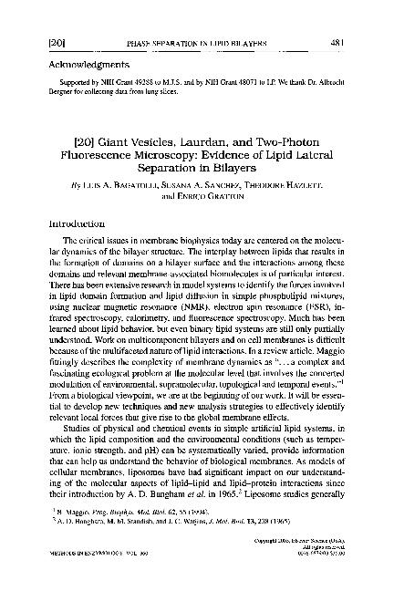
Giant vesicles, laurdan, and two-photon fluorescence microscopy: Evidence of lipid lateral separation in bilayers
Fecha de publicación:
03/2003
Editorial:
Elsevier Academic Press Inc.
Revista:
Methods In Enzymology
ISSN:
0076-6879
Idioma:
Inglés
Tipo de recurso:
Artículo publicado
Clasificación temática:
Resumen
This chapter describes giant vesicles, Laurdan, and two-photon fluorescence microscopy. The combination of Laurdan, giant unilamellar vesicles (GUVs), and two-photon fluorescence microscopy has been extremely useful in producing a microscopic picture of lipid-phase coexistence in the GUV bilayer model system. Laurdan is a unique probe, giving simultaneous information about morphology and phase state of lipid domains from fluorescence images. The critical issues in membrane biophysics today are centered on the molecular dynamics of the bilayer structure. The interplay between lipids that results in the formation of domains on a bilayer surface and the interactions among these domains and relevant membrane-associated biomolecules is of particular interest. The advantages of using a microscope as the optical arrangement are clear. The light collection efficiency of a well-designed microscope is greatly enhanced over other optical arrangements. In addition, the flexibility of fluorescence microscopes creates for the spectroscopist a malleable optical compartment that can be designed and readily redesigned as needed.
Archivos asociados
Licencia
Identificadores
Colecciones
Articulos(CIQUIBIC)
Articulos de CENTRO DE INVEST.EN QCA.BIOL.DE CORDOBA (P)
Articulos de CENTRO DE INVEST.EN QCA.BIOL.DE CORDOBA (P)
Citación
Bagatolli, Luis Alberto; Sanchez, Susana A.; Hazlett, Theodore; Gratton, Enrico; Giant vesicles, laurdan, and two-photon fluorescence microscopy: Evidence of lipid lateral separation in bilayers; Elsevier Academic Press Inc.; Methods In Enzymology; 360; 3-2003; 481-500
Compartir
Altmétricas


