Artículo
Expanding Actin Rings Zipper the Mouse Embryo for Blastocyst Formation
Zenker, Jennifer; White, Melanie D.; Gasnier, Maxime; Alvarez, Yanina Daniela ; Lim, Hui Yi Grace; Bissiere, Stephanie; Biro, Maté; Plachta, Nicolas
; Lim, Hui Yi Grace; Bissiere, Stephanie; Biro, Maté; Plachta, Nicolas
Fecha de publicación:
04/2018
Editorial:
Cell Press
Revista:
Cell
ISSN:
0092-8674
Idioma:
Inglés
Tipo de recurso:
Artículo publicado
Clasificación temática:
Resumen
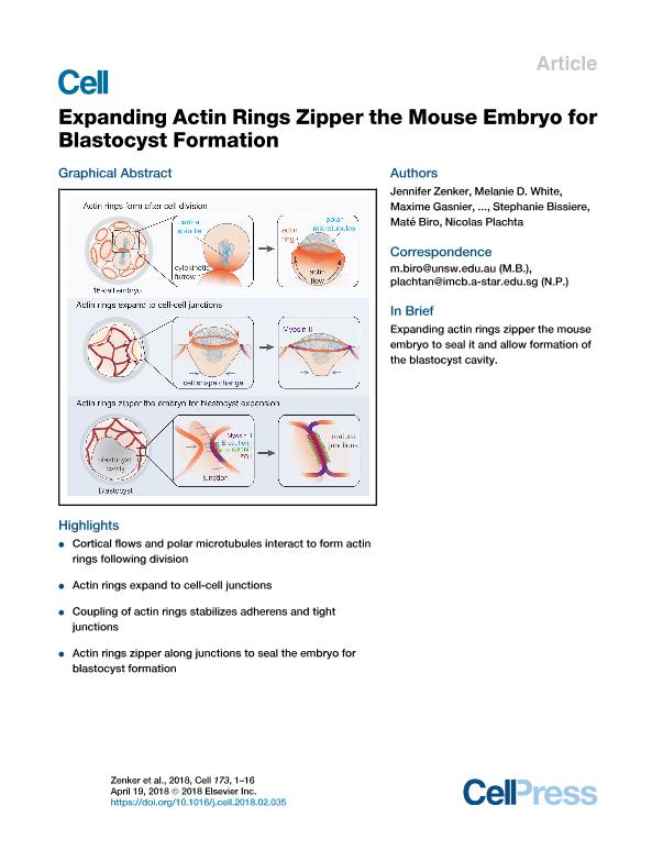
Transformation from morula to blastocyst is a defining event of preimplantation embryo development. During this transition, the embryo must establish a paracellular permeability barrier to enable expansion of the blastocyst cavity. Here, using live imaging of mouse embryos, we reveal an actin-zippering mechanism driving this embryo sealing. Preceding blastocyst stage, a cortical F-actin ring assembles at the apical pole of the embryo's outer cells. The ring structure forms when cortical actin flows encounter a network of polar microtubules that exclude F-actin. Unlike stereotypical actin rings, the actin rings of the mouse embryo are not contractile, but instead, they expand to the cell-cell junctions. Here, they couple to the junctions by recruiting and stabilizing adherens and tight junction components. Coupling of the actin rings triggers localized myosin II accumulation, and it initiates a tension-dependent zippering mechanism along the junctions that is required to seal the embryo for blastocyst formation. Expanding actin rings zipper the mouse embryo to seal it and allow formation of the blastocyst cavity.
Archivos asociados
Tamaño:
16.50Mb
Formato:
PDF
Licencia
 Excepto donde se diga explícitamente, este item se publica bajo la siguiente descripción:
Creative Commons Attribution-NonCommercial-ShareAlike 2.5 Unported (CC BY-NC-SA 2.5)
Excepto donde se diga explícitamente, este item se publica bajo la siguiente descripción:
Creative Commons Attribution-NonCommercial-ShareAlike 2.5 Unported (CC BY-NC-SA 2.5)
Identificadores
Colecciones
Articulos(IQUIBICEN)
Articulos de INSTITUTO DE QUIMICA BIOLOGICA DE LA FACULTAD DE CS. EXACTAS Y NATURALES
Articulos de INSTITUTO DE QUIMICA BIOLOGICA DE LA FACULTAD DE CS. EXACTAS Y NATURALES
Citación
Zenker, Jennifer; White, Melanie D.; Gasnier, Maxime; Alvarez, Yanina Daniela; Lim, Hui Yi Grace; et al.; Expanding Actin Rings Zipper the Mouse Embryo for Blastocyst Formation; Cell Press; Cell; 173; 3; 4-2018; 776-791.e17
Compartir


Altmétricas