Mostrar el registro sencillo del ítem
dc.contributor.author
Soriano, Silvia Viviana

dc.contributor.author
Debiaggi, Maria Florencia

dc.contributor.author
Pierangeli, Nora Beatriz
dc.contributor.author
Pianciola, Luis Alfredo
dc.contributor.author
Bergagna, Héctor Fabián Jesús
dc.contributor.author
Lazzarini, Lorena Evelina
dc.contributor.author
Mazzeo, Melina
dc.contributor.author
Basualdo Farjat, Juan Angel

dc.date.available
2019-07-02T21:13:43Z
dc.date.issued
2016-09
dc.identifier.citation
Soriano, Silvia Viviana; Debiaggi, Maria Florencia; Pierangeli, Nora Beatriz; Pianciola, Luis Alfredo; Bergagna, Héctor Fabián Jesús; et al.; First study about the development of adult Echinococcus canadensis G6 genotype of goat origin in experimentally infected dogs; Elsevier Science; Veterinary Parasitology; 228; 9-2016; 6-12
dc.identifier.issn
0304-4017
dc.identifier.uri
http://hdl.handle.net/11336/79044
dc.description.abstract
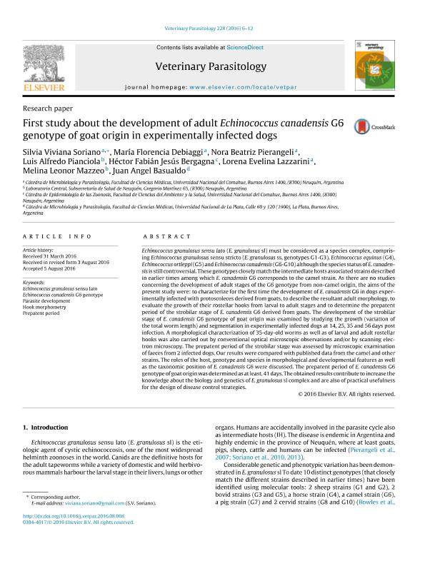
Echinococcus granulosus sensu lato (E. granulosus sl) must be considered as a species complex, comprising Echinococcus granulosus sensu stricto (E. granulosus ss, genotypes G1-G3), Echinococcus equinus (G4), Echinococcus ortleppi (G5) and Echinococcus canadensis (G6-G10) although the species status of E. canadensis is still controversial. These genotypes closely match the intermediate hosts associated strains described in earlier times among which E. canadensis G6 corresponds to the camel strain. As there are no studies concerning the development of adult stages of the G6 genotype from non-camel origin, the aims of the present study were: to characterize for the first time the development of E. canadensis G6 in dogs experimentally infected with protoscoleces derived from goats, to describe the resultant adult morphology, to evaluate the growth of their rostellar hooks from larval to adult stages and to determine the prepatent period of the strobilar stage of E. canadensis G6 derived from goats. The development of the strobilar stage of E. canadensis G6 genotype of goat origin was examined by studying the growth (variation of the total worm length) and segmentation in experimentally infected dogs at 14, 25, 35 and 56 days post infection. A morphological characterization of 35-day-old worms as well as of larval and adult rostellar hooks was also carried out by conventional optical microscopic observations and/or by scanning electron microscopy. The prepatent period of the strobilar stage was assessed by microscopic examination of faeces from 2 infected dogs. Our results were compared with published data from the camel and other strains. The roles of the host, genotype and species in morphological and developmental features as well as the taxonomic position of E. canadensis G6 were discussed. The prepatent period of E. canadensis G6 genotype of goat origin was determined as at least, 41 days. The obtained results contribute to increase the knowledge about the biology and genetics of E. granulosus sl complex and are also of practical usefulness for the design of disease control strategies.
dc.format
application/pdf
dc.language.iso
eng
dc.publisher
Elsevier Science

dc.rights
info:eu-repo/semantics/openAccess
dc.rights.uri
https://creativecommons.org/licenses/by-nc-sa/2.5/ar/
dc.subject
Echinococcus Canadensis G6 Genotype
dc.subject
Echinococcus Granulosus Sensu Lato
dc.subject
Hook Morphometry
dc.subject
Parasite Development
dc.subject
Prepatent Period
dc.subject.classification
Parasitología

dc.subject.classification
Ciencias de la Salud

dc.subject.classification
CIENCIAS MÉDICAS Y DE LA SALUD

dc.title
First study about the development of adult Echinococcus canadensis G6 genotype of goat origin in experimentally infected dogs
dc.type
info:eu-repo/semantics/article
dc.type
info:ar-repo/semantics/artículo
dc.type
info:eu-repo/semantics/publishedVersion
dc.date.updated
2019-06-11T15:22:00Z
dc.journal.volume
228
dc.journal.pagination
6-12
dc.journal.pais
Países Bajos

dc.journal.ciudad
Amsterdam
dc.description.fil
Fil: Soriano, Silvia Viviana. Universidad Nacional del Comahue. Facultad de Medicina; Argentina
dc.description.fil
Fil: Debiaggi, Maria Florencia. Consejo Nacional de Investigaciones Científicas y Técnicas. Centro Científico Tecnológico Conicet - Patagonia Norte; Argentina. Universidad Nacional del Comahue. Facultad de Medicina; Argentina
dc.description.fil
Fil: Pierangeli, Nora Beatriz. Universidad Nacional del Comahue. Facultad de Medicina; Argentina
dc.description.fil
Fil: Pianciola, Luis Alfredo. Provincia de Neuquén. Subsecretaría de Salud; Argentina
dc.description.fil
Fil: Bergagna, Héctor Fabián Jesús. Universidad Nacional del Comahue. Facultad de Medicina; Argentina
dc.description.fil
Fil: Lazzarini, Lorena Evelina. Universidad Nacional del Comahue. Facultad de Medicina; Argentina
dc.description.fil
Fil: Mazzeo, Melina. Provincia de Neuquén. Subsecretaría de Salud; Argentina
dc.description.fil
Fil: Basualdo Farjat, Juan Angel. Universidad Nacional de La Plata. Facultad de Ciencias Médicas; Argentina
dc.journal.title
Veterinary Parasitology

dc.relation.alternativeid
info:eu-repo/semantics/altIdentifier/url/https://www.ncbi.nlm.nih.gov/pubmed/27692331
dc.relation.alternativeid
info:eu-repo/semantics/altIdentifier/doi/http://dx.doi.org/10.1016/j.vetpar.2016.08.008
Archivos asociados
