Artículo
Meagre effects of disuse on the human fibula are not explained by bone size or geometry
Ireland, Alex; Capozza, Ricardo Francisco ; Cointry, Gustavo Roberto
 ; Nocciolino, Laura Marcela
 ; Ferretti, Jose Luis
 ; Rittweger, J.
 ; Cointry, Gustavo Roberto
 ; Nocciolino, Laura Marcela
 ; Ferretti, Jose Luis
 ; Rittweger, J.
Fecha de publicación:
02/2017
Editorial:
Springer London Ltd
Revista:
Osteoporosis International
ISSN:
0937-941X
e-ISSN:
1433-2965
Idioma:
Inglés
Tipo de recurso:
Artículo publicado
Clasificación temática:
Resumen
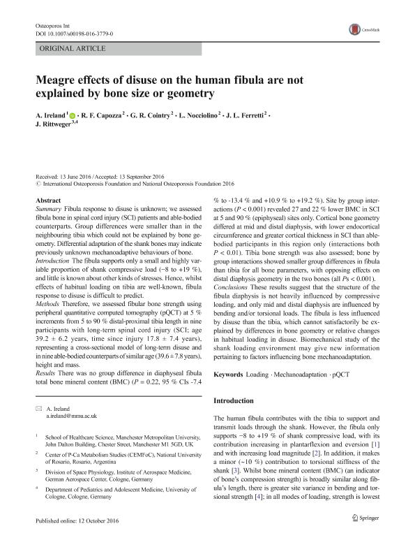
Summary: Fibula response to disuse is unknown; we assessed fibula bone in spinal cord injury (SCI) patients and able-bodied counterparts. Group differences were smaller than in the neighbouring tibia which could not be explained by bone geometry. Differential adaptation of the shank bones may indicate previously unknown mechanoadaptive behaviours of bone. Introduction: The fibula supports only a small and highly variable proportion of shank compressive load (−8 to +19 %), and little is known about other kinds of stresses. Hence, whilst effects of habitual loading on tibia are well-known, fibula response to disuse is difficult to predict. Methods: Therefore, we assessed fibular bone strength using peripheral quantitative computed tomography (pQCT) at 5 % increments from 5 to 90 % distal-proximal tibia length in nine participants with long-term spinal cord injury (SCI; age 39.2 ± 6.2 years, time since injury 17.8 ± 7.4 years), representing a cross-sectional model of long-term disuse and in nine able-bodied counterparts of similar age (39.6 ± 7.8 years), height and mass. Results: There was no group difference in diaphyseal fibula total bone mineral content (BMC) (P = 0.22, 95 % CIs -7.4 % to -13.4 % and +10.9 % to +19.2 %). Site by group interactions (P < 0.001) revealed 27 and 22 % lower BMC in SCI at 5 and 90 % (epiphyseal) sites only. Cortical bone geometry differed at mid and distal diaphysis, with lower endocortical circumference and greater cortical thickness in SCI than able-bodied participants in this region only (interactions both P < 0.01). Tibia bone strength was also assessed; bone by group interactions showed smaller group differences in fibula than tibia for all bone parameters, with opposing effects on distal diaphysis geometry in the two bones (all Ps < 0.001). Conclusions: These results suggest that the structure of the fibula diaphysis is not heavily influenced by compressive loading, and only mid and distal diaphysis are influenced by bending and/or torsional loads. The fibula is less influenced by disuse than the tibia, which cannot satisfactorily be explained by differences in bone geometry or relative changes in habitual loading in disuse. Biomechanical study of the shank loading environment may give new information pertaining to factors influencing bone mechanoadaptation.
Palabras clave:
Loading
,
Mechanoadaptation
,
Pqct
Archivos asociados
Licencia
Identificadores
Colecciones
Articulos(CCT - ROSARIO)
Articulos de CTRO.CIENTIFICO TECNOL.CONICET - ROSARIO
Articulos de CTRO.CIENTIFICO TECNOL.CONICET - ROSARIO
Citación
Ireland, Alex; Capozza, Ricardo Francisco; Cointry, Gustavo Roberto; Nocciolino, Laura Marcela; Ferretti, Jose Luis; et al.; Meagre effects of disuse on the human fibula are not explained by bone size or geometry; Springer London Ltd; Osteoporosis International; 28; 2; 2-2017; 633-641
Compartir
Altmétricas


