Mostrar el registro sencillo del ítem
dc.contributor.author
Berner, Silvia Inés
dc.contributor.author
de Bonis, Cristian
dc.contributor.author
Martinez, Diego
dc.contributor.author
Demarchi, Gianina

dc.contributor.author
Pérez Millán, María Inés

dc.contributor.author
Becu, Damasia

dc.contributor.author
Cristina, Silvia Carolina

dc.date.available
2016-08-30T18:05:20Z
dc.date.issued
2013-03
dc.identifier.citation
Berner, Silvia Inés; de Bonis, Cristian; Martinez, Diego; Demarchi, Gianina; Pérez Millán, María Inés; et al.; Mecanismos de tumorogenesis hipofisaria. Estudio de factores angiogénicos y de proliferación; Sociedad Argentina de Endocrinología y Metabolismo; Revista Argentina de Endocrinologia y Metabolismo; 50; 1; 3-2013; 19-24
dc.identifier.issn
0080-2077
dc.identifier.uri
http://hdl.handle.net/11336/7355
dc.description.abstract
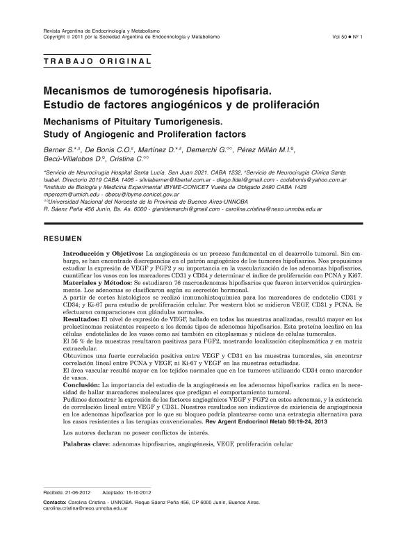
Introduction and objectives: Angiogenesis is an essential process in tumor development. Nevertheless, discrepancies in the angiogenic pattern of pituitary tumors, in terms of hormonal phenotype, size or invasiveness have been found. Our aim was to study the expression of VEGF and FGF2 growth factors, and their importance in the vascularization of pituitary adenomas. We also quantified blood vessels with the endothelial cell markers CD31 and CD34 determining the vascular area, and the proliferation rate through PCNA and Ki67 index. Materials and Methods: We studied 76 pituitary macroadenomas that were surgically resected in the period between 2006 and 2010 from a total of 276 patients with this pathology. Adenomas were classified into prolactinomas (PRL), somatotropinomas (GH), corticotropinomas (ACTH), non-functioning (NF) and plurihormonal (Ph) according to their hormonal secretion. Samples were collected in formalin, embedded in paraffin, and immunohistochemistry was performed from histological sections for endothelial markers CD31 and CD34; and for Ki-67 to study cell proliferation. VEGF, CD31 and PCNA were measured by Western blot. We compared results with normal glands (N=6). Results: VEGF expression levels, found in all of the samples analyzed, were higher in resistant prolactinomas than in other pituitary adenomas. This protein was detected in endothelial cells of blood vessels and in tumor cells cytoplasms and nuclei. Fifty-six percent of samples were positive for FGF2, the other potent angiogenic factor studied, showing cytoplasmatic and extracellular matrix localization. We obtained a strong positive correlation between VEGF and CD31 in tumor samples, but we did not find lineal correlation between PCNA and VEGF, or between Ki-67 and VEGF in the samples studied. The vascular area was higher in normal tissues than in tumors when CD34 was used as endothelial cell marker. Conclussion: The importance of studying angiogenesis in pituitary adenomas lies in the need to find molecular markers that can predict tumor behavior. We could demonstrate the expression of VEGF and FGF2, two potent angiogenic factors, and the existence of linear correlation between VEGF and CD31. Our results are indicative of the existence of angiogenesis in pituitary adenomas; therefore the blockage of angiogenesis might be proposed as an alternative strategy for cases of resistance to standard therapy.
dc.format
application/pdf
dc.language.iso
spa
dc.publisher
Sociedad Argentina de Endocrinología y Metabolismo

dc.rights
info:eu-repo/semantics/openAccess
dc.rights.uri
https://creativecommons.org/licenses/by-nc-sa/2.5/ar/
dc.subject
Adenomas Hipofisarios
dc.subject
Angiogenesis
dc.subject
Vegf
dc.subject
Proliferacion Celular
dc.subject.classification
Patología

dc.subject.classification
Medicina Básica

dc.subject.classification
CIENCIAS MÉDICAS Y DE LA SALUD

dc.title
Mecanismos de tumorogenesis hipofisaria. Estudio de factores angiogénicos y de proliferación
dc.title
Mechanisms of Pituitary Tumorigenesis. Study of Angiogenic and Proliferation factors
dc.type
info:eu-repo/semantics/article
dc.type
info:ar-repo/semantics/artículo
dc.type
info:eu-repo/semantics/publishedVersion
dc.date.updated
2016-05-03T13:44:22Z
dc.journal.volume
50
dc.journal.number
1
dc.journal.pagination
19-24
dc.journal.pais
Argentina

dc.journal.ciudad
Buenos Aires
dc.description.fil
Fil: Berner, Silvia Inés. Hospital Santa Lucía; Argentina. Clínica Santa Isabel; Argentina
dc.description.fil
Fil: de Bonis, Cristian. Clínica Santa Isabel; Argentina
dc.description.fil
Fil: Martinez, Diego. Hospital Santa Lucía; Argentina. Clínica Santa Isabel; Argentina
dc.description.fil
Fil: Demarchi, Gianina. Universidad Nacional del Noroeste de la Provincia de Buenos Aires. Departamento de Ciencias Básicas y Experimentales; Argentina
dc.description.fil
Fil: Pérez Millán, María Inés. Consejo Nacional de Investigaciones Científicas y Técnicas. Instituto de Biología y Medicina Experimental (i); Argentina
dc.description.fil
Fil: Becu, Damasia. Consejo Nacional de Investigaciones Científicas y Técnicas. Instituto de Biología y Medicina Experimental (i); Argentina
dc.description.fil
Fil: Cristina, Silvia Carolina. Universidad Nacional del Noroeste de la Provincia de Buenos Aires. Departamento de Ciencias Básicas y Experimentales; Argentina
dc.journal.title
Revista Argentina de Endocrinologia y Metabolismo

dc.relation.alternativeid
info:eu-repo/semantics/altIdentifier/url/http://www.raem.org.ar/numeros/2013-vol50/numero-01/vol50-01-002-eng.html
dc.relation.alternativeid
info:eu-repo/semantics/altIdentifier/url/http://www.scielo.org.ar/scielo.php?script=sci_arttext&pid=S1851-30342013000100002
Archivos asociados
