Mostrar el registro sencillo del ítem
dc.contributor.author
Caballero, Julieta N.
dc.contributor.author
Gervasi, Maria Gracia

dc.contributor.author
Veiga, Maria Florencia

dc.contributor.author
Dalvit, Gabriel Carlos

dc.contributor.author
Perez Martinez, Silvina Laura

dc.contributor.author
Cetica, Pablo Daniel

dc.contributor.author
Vazquez, Monica Hebe

dc.date.available
2016-07-18T19:12:05Z
dc.date.issued
2014-06
dc.identifier.citation
Caballero, Julieta N.; Gervasi, Maria Gracia; Veiga, Maria Florencia; Dalvit, Gabriel Carlos; Perez Martinez, Silvina Laura; et al.; Epithelial cadherin is present in bovine oviduct epithelial cells and gametes, and is involved in fertilization-related events; Elsevier; Theriogenology; 81; 9; 6-2014; 1189-1206
dc.identifier.issn
0093-691X
dc.identifier.uri
http://hdl.handle.net/11336/6572
dc.description.abstract
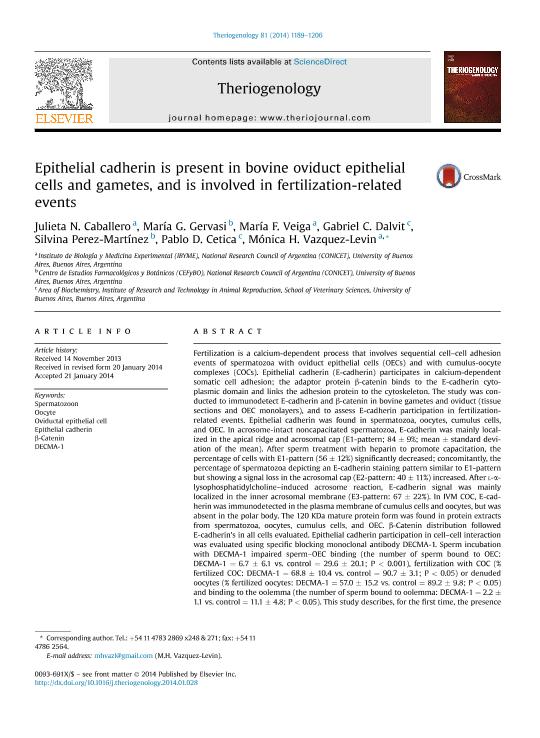
Fertilization is a calcium-dependent process that involves sequential cell-cell adhesion events of spermatozoa with oviduct epithelial cells (OECs) and with cumulus-oocyte complexes (COCs). Epithelial cadherin (E-cadherin) participates in calcium-dependent somatic cell adhesion; the adaptor protein β-catenin binds to the E-cadherin cytoplasmic domain and links the adhesion protein to the cytoskeleton. The study was conducted to immunodetect E-cadherin and β-catenin in bovine gametes and oviduct (tissue sections and OEC monolayers), and to assess E-cadherin participation in fertilization-related events. Epithelial cadherin was found in spermatozoa, oocytes, cumulus cells, and OEC. In acrosome-intact noncapacitated spermatozoa, E-cadherin was mainly localized in the apical ridge and acrosomal cap (E1-pattern; 84 ± 9%; mean ± standard deviation of the mean). After sperm treatment with heparin to promote capacitation, the percentage of cells with E1-pattern (56 ± 12%) significantly decreased; concomitantly, the percentage of spermatozoa depicting an E-cadherin staining pattern similar to E1-pattern but showing a signal loss in the acrosomal cap (E2-pattern: 40 ± 11%) increased. After l-α-lysophosphatidylcholine-induced acrosome reaction, E-cadherin signal was mainly localized in the inner acrosomal membrane (E3-pattern: 67 ± 22%). In IVM COC, E-cadherin was immunodetected in the plasma membrane of cumulus cells and oocytes, but was absent in the polar body. The 120 KDa mature protein form was found in protein extracts from spermatozoa, oocytes, cumulus cells, and OEC. β-Catenin distribution followed E-cadherin´s in all cells evaluated. Epithelial cadherin participation in cell-cell interaction was evaluated using specific blocking monoclonal antibody DECMA-1. Sperm incubation with DECMA-1 impaired sperm-OEC binding (the number of sperm bound to OEC: DECMA-1 = 6.7 ± 6.1 vs. control = 29.6 ± 20.1; P < 0.001), fertilization with COC (% fertilized COC: DECMA-1 = 68.8 ± 10.4 vs. control = 90.7 ± 3.1; P < 0.05) or denuded oocytes (% fertilized oocytes: DECMA-1 = 57.0 ± 15.2 vs. control = 89.2 ± 9.8; P < 0.05) and binding to the oolemma (the number of sperm bound to oolemma: DECMA-1 = 2.2 ± 1.1 vs. control = 11.1 ± 4.8; P < 0.05). This study describes, for the first time, the presence of E-cadherin in bovine spermatozoa, COC, and OEC, and shows evidence of its participation in sperm interaction with the oviduct and the oocyte during fertilization.
dc.format
application/pdf
dc.language.iso
eng
dc.publisher
Elsevier

dc.rights
info:eu-repo/semantics/openAccess
dc.rights.uri
https://creativecommons.org/licenses/by-nc-nd/2.5/ar/
dc.subject
Fertilization
dc.subject
Bovine
dc.subject
Epithelial Cadherin
dc.subject
Gamete Adhesion
dc.subject.classification
Biología Reproductiva

dc.subject.classification
Ciencias Biológicas

dc.subject.classification
CIENCIAS NATURALES Y EXACTAS

dc.title
Epithelial cadherin is present in bovine oviduct epithelial cells and gametes, and is involved in fertilization-related events
dc.type
info:eu-repo/semantics/article
dc.type
info:ar-repo/semantics/artículo
dc.type
info:eu-repo/semantics/publishedVersion
dc.date.updated
2016-06-15T19:10:13Z
dc.journal.volume
81
dc.journal.number
9
dc.journal.pagination
1189-1206
dc.journal.pais
Países Bajos

dc.journal.ciudad
Amsterdam
dc.description.fil
Fil: Caballero, Julieta N.. Consejo Nacional de Investigaciones Científicas y Técnicas. Instituto de Biología y Medicina Experimental (i); Argentina
dc.description.fil
Fil: Gervasi, Maria Gracia. Consejo Nacional de Investigaciones Científicas y Técnicas. Oficina de Coordinación Administrativa Houssay. Centro de Estudios Farmacológicos y Botánicos; Argentina
dc.description.fil
Fil: Veiga, Maria Florencia. Consejo Nacional de Investigaciones Científicas y Técnicas. Instituto de Biología y Medicina Experimental (i); Argentina
dc.description.fil
Fil: Dalvit, Gabriel Carlos. Universidad de Buenos Aires. Facultad de Ciencias Veterinarias; Argentina
dc.description.fil
Fil: Perez Martinez, Silvina Laura. Consejo Nacional de Investigaciones Científicas y Técnicas. Oficina de Coordinación Administrativa Houssay. Centro de Estudios Farmacológicos y Botánicos; Argentina
dc.description.fil
Fil: Cetica, Pablo Daniel. Universidad de Buenos Aires. Facultad de Ciencias Veterinarias; Argentina. Consejo Nacional de Investigaciones Científicas y Técnicas; Argentina
dc.description.fil
Fil: Vazquez, Monica Hebe. Consejo Nacional de Investigaciones Científicas y Técnicas. Instituto de Biología y Medicina Experimental (i); Argentina
dc.journal.title
Theriogenology

dc.relation.alternativeid
info:eu-repo/semantics/altIdentifier/url/http://www.sciencedirect.com/science/article/pii/S0093691X14000612
dc.relation.alternativeid
info:eu-repo/semantics/altIdentifier/doi/http://dx.doi.org/10.1016/j.theriogenology.2014.01.028
Archivos asociados
