Artículo
Apoptosis and cell proliferation in porcine placental vascularization
Sanchis, Eva Gabriela ; Cristofolini, Andrea Lorena
 ; Fiorimanti, Mariana Rita
 ; Barbeito, Claudio Gustavo
 ; Merkis, Cecilia Inés
 ; Cristofolini, Andrea Lorena
 ; Fiorimanti, Mariana Rita
 ; Barbeito, Claudio Gustavo
 ; Merkis, Cecilia Inés
Fecha de publicación:
09/2017
Editorial:
Elsevier Science
Revista:
Animal Reproduction Science
ISSN:
0378-4320
Idioma:
Inglés
Tipo de recurso:
Artículo publicado
Clasificación temática:
Resumen
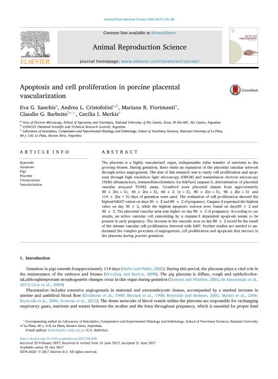
The placenta is a highly vascularized organ, indispensable tothe transfer of nutrients to the growing fetuses. During gestation, there exists an expansion of the placental vascular network through active angiogenesis. The aim of this research was to study cell proliferation and apoptosis through high resolution light microscopy (HRLM) and transmission electron microscopy (TEM) ultrastructure, immunohistochemistry for Ki67and caspase-3, determination of placental vascular area,and TUNEL assay. Crossbred sows placental tissues from approximately 30 ± 2(n = 5), 40 ± 2(n = 5), 60 ± 2 (n = 5), 80 ± 2(n = 5), 90 ± 2(n = 5) and 114 ± 2(n = 5) days of gestation were used. The evaluation of cell proliferation showed the highest%Ki67 values on days 30 ± 2 and 80 ± 2 of pregnancy. Caspase-3 expressed the highest value on day 30 ± 2, while the highest apoptotic indexes were found on days30 ± 2 and 90 ± 2. The placental vascular area was higher on day 80 ± 2 of pregnancy. According to our results, an active vascular cell remodeling by a caspase-3 dependent apoptosis seems to be present in early pregnancy. The increase in the vascular area on day 80 ± 2 would be the result of the intense vascular cell proliferation detected with Ki67. Further studies are needed to understand the complex processes of angiogenesis, cell proliferation and apoptosis that interact in the placenta during porcine gestation.
Palabras clave:
Apoptosis
,
Pigs
,
Placenta
,
Ultrastructure
,
Vascularization
Archivos asociados
Licencia
Identificadores
Colecciones
Articulos(CCT - LA PLATA)
Articulos de CTRO.CIENTIFICO TECNOL.CONICET - LA PLATA
Articulos de CTRO.CIENTIFICO TECNOL.CONICET - LA PLATA
Citación
Sanchis, Eva Gabriela; Cristofolini, Andrea Lorena; Fiorimanti, Mariana Rita; Barbeito, Claudio Gustavo; Merkis, Cecilia Inés; Apoptosis and cell proliferation in porcine placental vascularization; Elsevier Science; Animal Reproduction Science; 184; 9-2017; 20-28
Compartir
Altmétricas


