Artículo
Estrogen receptor beta growth inhibitory effects are repressed through activation of MAPK and PI3K signalling in mammary epithelial and breast cancer cells
Cotrim, C. Z.; Fabris, Victoria Teresa ; Doria, M. L.; Lindberg, K.; Gustafsson, J. A.; Amado, F.; Lanari, Claudia Lee Malvina
 ; Helguero, L. A.
 ; Doria, M. L.; Lindberg, K.; Gustafsson, J. A.; Amado, F.; Lanari, Claudia Lee Malvina
 ; Helguero, L. A.
Fecha de publicación:
09/05/2013
Editorial:
Nature Publishing Group
Revista:
Oncogene
ISSN:
0950-9232
e-ISSN:
1476-5594
Idioma:
Inglés
Tipo de recurso:
Artículo publicado
Clasificación temática:
Resumen
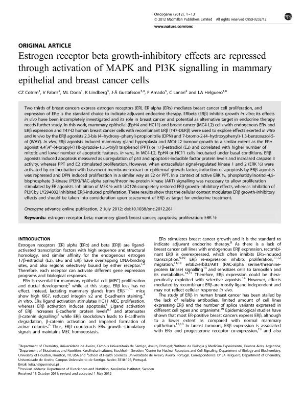
Two thirds of breast cancers express estrogen receptors (ER). ER alpha (ERa) mediates breast cancer cell proliferation, and expression of ERa is the standard choice to indicate adjuvant endocrine therapy. ERbeta (ERb) inhibits growth in vitro; its effects in vivo have been incompletely investigated and its role in breast cancer and potential as alternative target in endocrine therapy needs further study. In this work, mammary epithelial (EpH4 and HC11) and breast cancer (MC4-L2) cells with endogenous ERa and ERb expression and T47-D human breast cancer cells with recombinant ERb (T47-DERb) were used to explore effects exerted in vitro and in vivo by the ERb agonists 2,3-bis (4?hydroxy?phenyl)-propionitrile (DPN) and 7-bromo-2-(4?hydroxyphenyl)-1,3-benzoxazol-5-ol (WAY). In vivo, ERb agonists induced mammary gland hyperplasia and MC4-L2 tumour growth to a similar extent as the Era agonist 4,4´,4´´-(4-propyl-(1H)-pyrazole-1,3,5-triyl) trisphenol (PPT) or 17b-estradiol (E2) and correlated with higher number of mitotic and lower number of apoptotic features. In vitro, in MC4-L2, EpH4 or HC11 cells incubated under basal conditions, ERb agonists induced apoptosis measured as upregulation of p53 and apoptosis-inducible factor protein levels and increased caspase 3 activity, whereas PPT and E2 stimulated proliferation. However, when extracellular signal-regulated kinase 1 and 2 (ERK ½) were activated by co-incubation with basement membrane extract or epidermal growth factor, induction of apoptosis by ERb agonists was repressed and DPN induced proliferation in a similar way as E2 or PPT. In a context of active ERK ½, phosphatidylinositol-4,5-bisphosphate 3-kinase (PI3K)/RAC-alpha serine/threonine-protein kinase (AKT) signalling was necessary to allow proliferation stimulated by ER agonists. Inhibition of MEK ½ with UO126 completely restored ERb growth-inhibitory effects, whereas inhibition of PI3K by LY294002 inhibited ERb-induced proliferation. These results show that the cellular context modulates ERb growth-inhibitory effects and should be taken into consideration upon assessment of ERb as target for endocrine treatment.
Archivos asociados
Licencia
Identificadores
Colecciones
Articulos(IBYME)
Articulos de INST.DE BIOLOGIA Y MEDICINA EXPERIMENTAL (I)
Articulos de INST.DE BIOLOGIA Y MEDICINA EXPERIMENTAL (I)
Citación
Cotrim, C. Z.; Fabris, Victoria Teresa; Doria, M. L.; Lindberg, K.; Gustafsson, J. A.; et al.; Estrogen receptor beta growth inhibitory effects are repressed through activation of MAPK and PI3K signalling in mammary epithelial and breast cancer cells; Nature Publishing Group; Oncogene; 32; 19; 9-5-2013; 2390-2402
Compartir
Altmétricas


