Mostrar el registro sencillo del ítem
dc.contributor.author
Crim, Nicolás

dc.contributor.author
Esposito, Evangelina

dc.contributor.author
Monti, Jose Rodolfo

dc.contributor.author
Correa, Leandro Javier

dc.contributor.author
Serra, Horacio Marcelo

dc.contributor.author
Urrets Zavalía, Julio Alberto

dc.date.available
2018-06-29T17:08:26Z
dc.date.issued
2017-12-01
dc.identifier.citation
Crim, Nicolás; Esposito, Evangelina; Monti, Jose Rodolfo; Correa, Leandro Javier; Serra, Horacio Marcelo; et al.; Myopia as a risk factor for subsequent retinal tears in the course of a symptomatic posterior vitreous detachment; BioMed Central; BMC Ophthalmology; 17; 1; 1-12-2017; 226-226
dc.identifier.issn
1471-2415
dc.identifier.uri
http://hdl.handle.net/11336/50670
dc.description.abstract
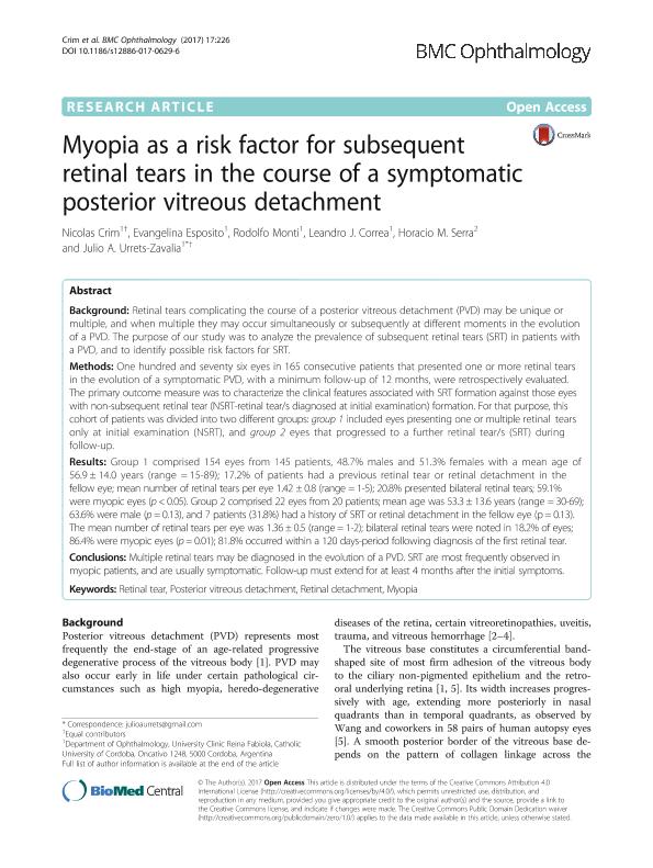
Background: Retinal tears complicating the course of a posterior vitreous detachment (PVD) may be unique or multiple, and when multiple they may occur simultaneously or subsequently at different moments in the evolution of a PVD. The purpose of our study was to analyze the prevalence of subsequent retinal tears (SRT) in patients with a PVD, and to identify possible risk factors for SRT. Methods: One hundred and seventy six eyes in 165 consecutive patients that presented one or more retinal tears in the evolution of a symptomatic PVD, with a minimum follow-up of 12 months, were retrospectively evaluated. The primary outcome measure was to characterize the clinical features associated with SRT formation against those eyes with non-subsequent retinal tear (NSRT-retinal tear/s diagnosed at initial examination) formation. For that purpose, this cohort of patients was divided into two different groups: group 1 included eyes presenting one or multiple retinal tears only at initial examination (NSRT), and group 2 eyes that progressed to a further retinal tear/s (SRT) during follow-up. Results: Group 1 comprised 154 eyes from 145 patients, 48.7% males and 51.3% females with a mean age of 56.9 ± 14.0 years (range = 15-89); 17.2% of patients had a previous retinal tear or retinal detachment in the fellow eye; mean number of retinal tears per eye 1.42 ± 0.8 (range = 1-5); 20.8% presented bilateral retinal tears; 59.1% were myopic eyes (p < 0.05). Group 2 comprised 22 eyes from 20 patients; mean age was 53.3 ± 13.6 years (range = 30-69); 63.6% were male (p = 0.13), and 7 patients (31.8%) had a history of SRT or retinal detachment in the fellow eye (p = 0.13). The mean number of retinal tears per eye was 1.36 ± 0.5 (range = 1-2); bilateral retinal tears were noted in 18.2% of eyes; 86.4% were myopic eyes (p = 0.01); 81.8% occurred within a 120 days-period following diagnosis of the first retinal tear. Conclusions: Multiple retinal tears may be diagnosed in the evolution of a PVD. SRT are most frequently observed in myopic patients, and are usually symptomatic. Follow-up must extend for at least 4 months after the initial symptoms.
dc.format
application/pdf
dc.language.iso
eng
dc.publisher
BioMed Central

dc.rights
info:eu-repo/semantics/openAccess
dc.rights.uri
https://creativecommons.org/licenses/by/2.5/ar/
dc.subject
Myopia
dc.subject
Posterior Vitreous Detachment
dc.subject
Retinal Detachment
dc.subject
Retinal Tear
dc.subject.classification
Medicina Critica y de Emergencia

dc.subject.classification
Medicina Clínica

dc.subject.classification
CIENCIAS MÉDICAS Y DE LA SALUD

dc.title
Myopia as a risk factor for subsequent retinal tears in the course of a symptomatic posterior vitreous detachment
dc.type
info:eu-repo/semantics/article
dc.type
info:ar-repo/semantics/artículo
dc.type
info:eu-repo/semantics/publishedVersion
dc.date.updated
2018-06-11T12:59:21Z
dc.journal.volume
17
dc.journal.number
1
dc.journal.pagination
226-226
dc.journal.pais
Reino Unido

dc.journal.ciudad
Londres
dc.description.fil
Fil: Crim, Nicolás. Universidad Catolica de Córdoba. Facultad de Medicina. Clinica Universitaria Reina Fabiola; Argentina
dc.description.fil
Fil: Esposito, Evangelina. Universidad Catolica de Córdoba. Facultad de Medicina. Clinica Universitaria Reina Fabiola; Argentina
dc.description.fil
Fil: Monti, Jose Rodolfo. Universidad Catolica de Córdoba. Facultad de Medicina. Clinica Universitaria Reina Fabiola; Argentina
dc.description.fil
Fil: Correa, Leandro Javier. Universidad Catolica de Córdoba. Facultad de Medicina. Clinica Universitaria Reina Fabiola; Argentina
dc.description.fil
Fil: Serra, Horacio Marcelo. Universidad Catolica de Córdoba. Facultad de Medicina. Clinica Universitaria Reina Fabiola; Argentina. Consejo Nacional de Investigaciones Científicas y Técnicas. Centro Científico Tecnológico Córdoba. Centro de Investigaciones en Bioquímica Clínica e Inmunología; Argentina
dc.description.fil
Fil: Urrets Zavalía, Julio Alberto. Universidad Catolica de Córdoba. Facultad de Medicina. Clinica Universitaria Reina Fabiola; Argentina
dc.journal.title
BMC Ophthalmology
dc.relation.alternativeid
info:eu-repo/semantics/altIdentifier/doi/http://dx.doi.org/10.1186/s12886-017-0629-6
dc.relation.alternativeid
info:eu-repo/semantics/altIdentifier/url/https://bmcophthalmol.biomedcentral.com/articles/10.1186/s12886-017-0629-6
Archivos asociados
