Artículo
Anatomical variations of the thoracic duct: a preliminary report in adults and fetal specimens
Fecha de publicación:
12/2016
Editorial:
International Society of Lymphology
Revista:
Lymphology
ISSN:
0024-7766
e-ISSN:
2522-7963
Idioma:
Inglés
Tipo de recurso:
Artículo publicado
Clasificación temática:
Resumen
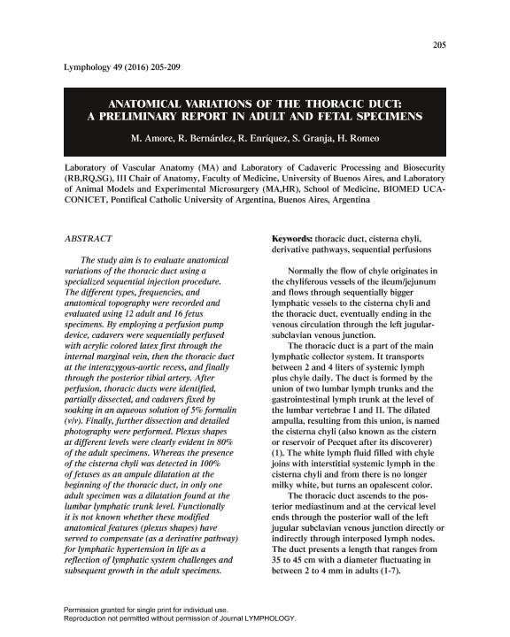
The study aim is to evaluate anatomical variations of the thoracic duct using a specialized sequential injection procedure. The different types, frequencies, and anatomical topography were recorded and evaluated using 12 adult and 16 fetus specimens. By employing a perfusion pump device, cadavers were sequentially perfused with acrylic colored latex first through the internal marginal vein, then the thoracic duct at the interazygous-aortic recess, and finally through the posterior tibial artery. After perfusion, thoracic ducts were identified, partially dissected, and cadavers fixed by soaking in an aqueous solution of 5% formalin (v/v). Finally, further dissection and detailed photography were performed. Plexus shapes at different levels were clearly evident in 80% of the adult specimens. Whereas the presence of the cisterna chyli was detected in 100% of fetuses as an ampule dilatation at the beginning of the thoracic duct, in only one adult specimen was a dilatation found at the lumbar lymphatic trunk level. Functionally it is not known whether these modified anatomical features (plexus shapes) have served to compensate (as a derivative pathway) for lymphatic hypertension in life as a reflection of lymphatic system challenges and subsequent growth in the adult specimens.
Palabras clave:
Thoracic Duct
,
Cisterna Chyli
,
Derivative Pathways
,
Sequential Perfusions
Archivos asociados
Licencia
Identificadores
Colecciones
Articulos(OCA HOUSSAY)
Articulos de OFICINA DE COORDINACION ADMINISTRATIVA HOUSSAY
Articulos de OFICINA DE COORDINACION ADMINISTRATIVA HOUSSAY
Citación
Amore, Miguel A.; Bernárdez, R.; Enríquez, R.; Granja, S.; Romeo, Horacio Eduardo; Anatomical variations of the thoracic duct: a preliminary report in adults and fetal specimens; International Society of Lymphology; Lymphology; 49; 4; 12-2016; 205-209
Compartir


