Mostrar el registro sencillo del ítem
dc.contributor.author
Ibañez Rodriguez, María Paula

dc.contributor.author
Noctor, Stephen C.
dc.contributor.author
Muñoz, Estela Maris

dc.date.available
2018-06-15T22:13:02Z
dc.date.issued
2016-11
dc.identifier.citation
Ibañez Rodriguez, María Paula; Noctor, Stephen C.; Muñoz, Estela Maris; Cellular basis of pineal gland development: Emerging role of microglia as phenotype regulator; Public Library of Science; Plos One; 11; 11; 11-2016; 1-24
dc.identifier.issn
1932-6203
dc.identifier.uri
http://hdl.handle.net/11336/48926
dc.description.abstract
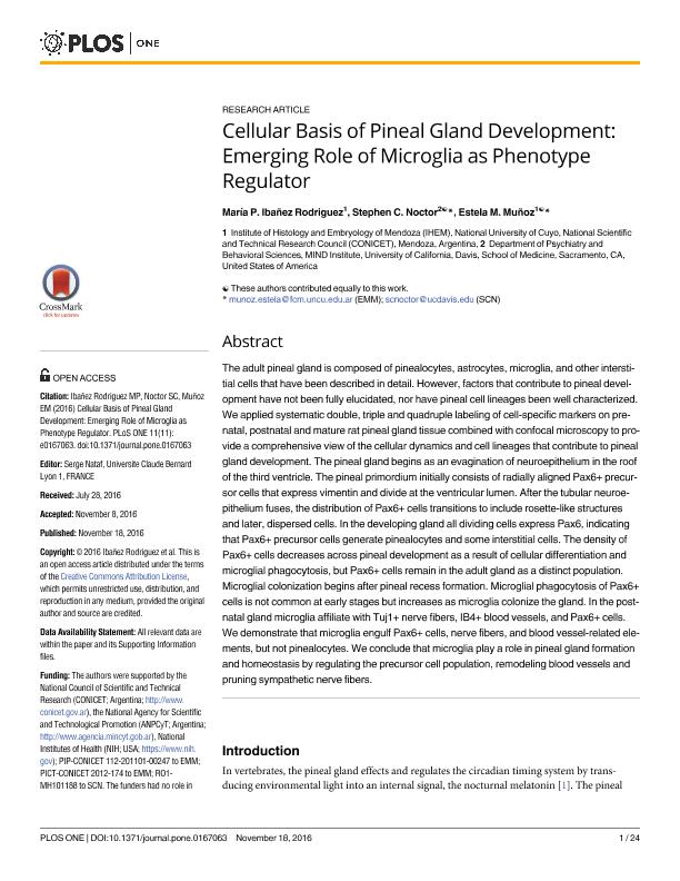
The adult pineal gland is composed of pinealocytes, astrocytes, microglia, and other interstitial cells that have been described in detail. However, factors that contribute to pineal development have not been fully elucidated, nor have pineal cell lineages been well characterized. We applied systematic double, triple and quadruple labeling of cell-specific markers on prenatal, postnatal and mature rat pineal gland tissue combined with confocal microscopy to provide a comprehensive view of the cellular dynamics and cell lineages that contribute to pineal gland development. The pineal gland begins as an evagination of neuroepithelium in the roof of the third ventricle. The pineal primordium initially consists of radially aligned Pax6+ precursor cells that express vimentin and divide at the ventricular lumen. After the tubular neuroepithelium fuses, the distribution of Pax6+ cells transitions to include rosette-like structures and later, dispersed cells. In the developing gland all dividing cells express Pax6, indicating that Pax6+ precursor cells generate pinealocytes and some interstitial cells. The density of Pax6+ cells decreases across pineal development as a result of cellular differentiation and microglial phagocytosis, but Pax6+ cells remain in the adult gland as a distinct population. Microglial colonization begins after pineal recess formation. Microglial phagocytosis of Pax6+ cells is not common at early stages but increases as microglia colonize the gland. In the postnatal gland microglia affiliate with Tuj1+ nerve fibers, IB4+ blood vessels, and Pax6+ cells. We demonstrate that microglia engulf Pax6+ cells, nerve fibers, and blood vessel-related elements, but not pinealocytes. We conclude that microglia play a role in pineal gland formation and homeostasis by regulating the precursor cell population, remodeling blood vessels and pruning sympathetic nerve fibers.
dc.format
application/pdf
dc.language.iso
eng
dc.publisher
Public Library of Science

dc.rights
info:eu-repo/semantics/openAccess
dc.rights.uri
https://creativecommons.org/licenses/by-nc-sa/2.5/ar/
dc.subject
Pineal Gland
dc.subject
Microglia
dc.subject
Precursor Cells
dc.subject
Iba1
dc.subject
Pax6
dc.subject.classification
Otras Ciencias Biológicas

dc.subject.classification
Ciencias Biológicas

dc.subject.classification
CIENCIAS NATURALES Y EXACTAS

dc.title
Cellular basis of pineal gland development: Emerging role of microglia as phenotype regulator
dc.type
info:eu-repo/semantics/article
dc.type
info:ar-repo/semantics/artículo
dc.type
info:eu-repo/semantics/publishedVersion
dc.date.updated
2018-06-13T16:52:47Z
dc.journal.volume
11
dc.journal.number
11
dc.journal.pagination
1-24
dc.journal.pais
Estados Unidos

dc.journal.ciudad
San Francisco
dc.description.fil
Fil: Ibañez Rodriguez, María Paula. Consejo Nacional de Investigaciones Científicas y Técnicas. Centro Científico Tecnológico Conicet - Mendoza. Instituto de Histología y Embriología de Mendoza Dr. Mario H. Burgos. Universidad Nacional de Cuyo. Facultad de Cienicas Médicas. Instituto de Histología y Embriología de Mendoza Dr. Mario H. Burgos; Argentina
dc.description.fil
Fil: Noctor, Stephen C.. University of California; Estados Unidos
dc.description.fil
Fil: Muñoz, Estela Maris. Consejo Nacional de Investigaciones Científicas y Técnicas. Centro Científico Tecnológico Conicet - Mendoza. Instituto de Histología y Embriología de Mendoza Dr. Mario H. Burgos. Universidad Nacional de Cuyo. Facultad de Cienicas Médicas. Instituto de Histología y Embriología de Mendoza Dr. Mario H. Burgos; Argentina
dc.journal.title
Plos One

dc.relation.alternativeid
info:eu-repo/semantics/altIdentifier/doi/http://dx.doi.org/10.1371/journal.pone.0167063
dc.relation.alternativeid
info:eu-repo/semantics/altIdentifier/url/http://journals.plos.org/plosone/article?id=10.1371/journal.pone.0167063
Archivos asociados
