Artículo
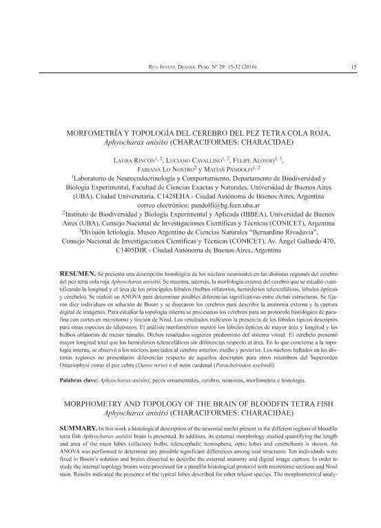
Se presenta una descripción histológica de los núcleos neuronales en las distintas regiones del cerebro del pez tetra cola roja Aphyocharax anisitsi. Se muestra, además, la morfología externa del cerebro que se estudió cuan-tificando la longitud y el área de los principales lóbulos (bulbos olfatorios, hemisferios telencefálicos, lóbulos ópticos y cerebelo). Se realizó un ANOVA para determinar posibles diferencias significativas entre dichas estructuras. Se fija-ron diez individuos en solución de Bouin y se disecaron los cerebros para describir la anatomía externa y la captura digital de imágenes. Para estudiar la topología interna se procesaron los cerebros para un protocolo histológico de para-fina con cortes en micrótomo y tinción de Nissl. Los resultados indicaron la presencia de los lóbulos típicos descriptos para otras especies de teleósteos. El análisis morfométrico mostró los lóbulos ópticos de mayor área y longitud y los bulbos olfatorios de menor tamaño. Dichos resultados sugieren predominio del sistema visual. El cerebelo presentó mayor longitud total que los hemisferios telencefálicos sin diferencias respecto al área. En lo que concierne a la topo-logía interna, se observó a los núcleos asociados al cerebro anterior, medio y posterior. Los núcleos hallados en las dis-tintas regiones no presentaron diferencias respecto de aquellos descriptos para otros miembros del Superorden Ostariophysi como el pez cebra (Danio rerio) o el neón cardenal (Paracheirodon axelrodi). In this work a histological description of the neuronal nuclei present in the different regions of bloodfin tetra fish Aphyocharax anisitsi brain is presented. In addition, its external morphology studied quantifying the length and area of the main lobes (olfactory bulbs, telencephalic hemisphere, optic lobes and cerebellum) is shown. An ANOVA was performed to determine any possible significant differences among said structures. Ten individuals were fixed in Bouin’s solution and brains dissected to describe the external anatomy and digital image capture. In order to study the internal topology brains were processed for a paraffin histological protocol with microtome sections and Nissl stain. Results indicated the presence of the typical lobes described for other teleost species. The morphometrical analy- sis showed the optic lobes of the largest area and length and the olfactory bulbs of the smallest size. Said results suggest a predominance of the visual system. The cerebellum presented a larger total length than the telencephalic hemispheres with no differences as regards the area. Concerning the internal topology, it was observed that nuclei were associated to the anterior, medium and posterior brain. The nuclei found in the different regions did not show differences with regard to those described for other members of the Ostariophysi Superorder such as the zebra fish (Danio rerio) or the cardinal tetra (Paracheirodon axelrodi).
Morfometría y topología del cerebro del pez tetra cola roja, aphyocharax anisitsi (characiformes: characidae)
Título:
Morphometry and topology of the brain of bloodfin tetra fish aphyocharax anisitsi (characiformes: characidae)
Rincón Camacho, Laura ; Cavallino, Luciano
 ; Alonso, Felipe
 ; Lo Nostro, Fabiana Laura
 ; Pandolfi, Matias
 ; Cavallino, Luciano
 ; Alonso, Felipe
 ; Lo Nostro, Fabiana Laura
 ; Pandolfi, Matias
Fecha de publicación:
12/2016
Editorial:
Instituto Nacional de Investigación y Desarrollo Pesquero
Revista:
Revista de Investigacion y Desarrollo Pesquero
ISSN:
0325-6375
Idioma:
Español
Tipo de recurso:
Artículo publicado
Clasificación temática:
Resumen
Palabras clave:
Aphyocharax Anisitsi
,
Peces Ornamentales
,
Cerebro
,
Neuronas
,
Morfometría
,
Histología
Archivos asociados
Licencia
Identificadores
Colecciones
Articulos(MACNBR)
Articulos de MUSEO ARG.DE CS.NAT "BERNARDINO RIVADAVIA"
Articulos de MUSEO ARG.DE CS.NAT "BERNARDINO RIVADAVIA"
Citación
Rincón Camacho, Laura; Cavallino, Luciano; Alonso, Felipe; Lo Nostro, Fabiana Laura; Pandolfi, Matias; Morfometría y topología del cerebro del pez tetra cola roja, aphyocharax anisitsi (characiformes: characidae); Instituto Nacional de Investigación y Desarrollo Pesquero; Revista de Investigacion y Desarrollo Pesquero; 29; 12-2016; 15-32
Compartir


