Artículo
A background Ca 2+ entry pathway mediated by TRPC1/TRPC4 is critical for development of pathological cardiac remodelling
Camacho Londoño, Juan E.; Tian, Qinghai; Hammer, Karin; Schröder, Laura; Camacho Londoño, Julia; Reil, Jan C.; He, Tao; Oberhofer, Martin; Mannebach, Stefanie; Mathar, Ilka; Philipp, Stephan E.; Tabellion, Wiebke; Schweda, Frank; Dietrich, Alexander; Kaestner, Lars; Laufs, Ulrich; Birnbaumer, Lutz ; Flockerzi, Veit; Freichel, Marc; Lipp, Peter
 ; Flockerzi, Veit; Freichel, Marc; Lipp, Peter
Fecha de publicación:
09/2015
Editorial:
Oxford University Press
Revista:
European Heart Journal
ISSN:
0195-668X
Idioma:
Inglés
Tipo de recurso:
Artículo publicado
Clasificación temática:
Resumen
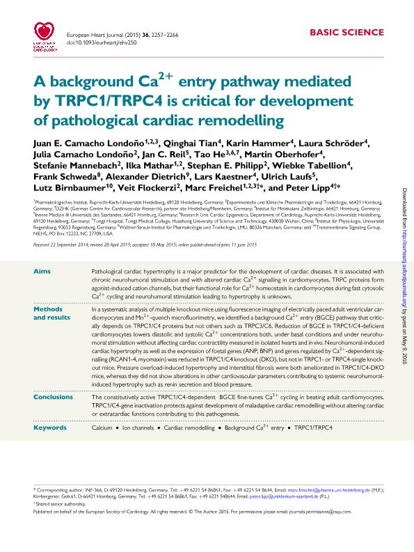
Aims: Pathological cardiac hypertrophy is a major predictor for the development of cardiac diseases. It is associated with chronic neurohumoral stimulation and with altered cardiac Ca2+ signalling in cardiomyocytes. TRPC proteins form agonist-induced cation channels, but their functional role for Ca2+ homeostasis in cardiomyocytes during fast cytosolic Ca2+ cycling and neurohumoral stimulation leading to hypertrophy is unknown. Methods and results: In a systematic analysis of multiple knockout mice using fluorescence imaging of electrically paced adult ventricular cardiomyocytes and Mn2+-quench microfluorimetry, we identified a background Ca2+ entry (BGCE) pathway that critically depends on TRPC1/C4 proteins but not others such as TRPC3/C6. Reduction of BGCE in TRPC1/C4-deficient cardiomyocytes lowers diastolic and systolic Ca2+ concentrations both, under basal conditions and under neurohumoral stimulation without affecting cardiac contractility measured in isolated hearts and in vivo. Neurohumoral-induced cardiac hypertrophy as well as the expression of foetal genes (ANP, BNP) and genes regulated by Ca2+-dependent signalling (RCAN1-4, myomaxin) was reduced in TRPC1/C4 knockout (DKO), but not in TRPC1- or TRPC4-single knockout mice. Pressure overload-induced hypertrophy and interstitial fibrosis were both ameliorated in TRPC1/C4-DKO mice, whereas they did not show alterations in other cardiovascular parameters contributing to systemic neurohumoral-induced hypertrophy such as renin secretion and blood pressure. Conclusions: The constitutively active TRPC1/C4-dependent BGCE fine-tunes Ca2+ cycling in beating adult cardiomyocytes. TRPC1/C4-gene inactivation protects against development of maladaptive cardiac remodelling without altering cardiac or extracardiac functions contributing to this pathogenesis.
Palabras clave:
Calcium
,
Ion Channels
,
Cardiac Remodelling
,
Background Ca2+ Entry
,
Trpc1/Trpc4
Archivos asociados
Licencia
Identificadores
Colecciones
Articulos(IIB-INTECH)
Articulos de INST.DE INVEST.BIOTECNOLOGICAS - INSTITUTO TECNOLOGICO CHASCOMUS
Articulos de INST.DE INVEST.BIOTECNOLOGICAS - INSTITUTO TECNOLOGICO CHASCOMUS
Citación
Camacho Londoño, Juan E.; Tian, Qinghai; Hammer, Karin; Schröder, Laura; Camacho Londoño, Julia; et al.; A background Ca 2+ entry pathway mediated by TRPC1/TRPC4 is critical for development of pathological cardiac remodelling; Oxford University Press; European Heart Journal; 36; 33; 9-2015; 2257-2266
Compartir
Altmétricas


