Artículo
Idiopathic atrophoderma of Pasini and Pierini: A case study of collagen and elastin texture by multiphoton microscopy
Vieira Damiani, Gislaine; Lage, Denise; Daldon, Patrícia Érica Christofoletti; Alves, Caroline Romanelli Tibúrcio; Cintra, Maria Letícia; Metze, Konradin; Adur, Javier Fernando ; Pelegati, Vitor B.; Carvalho, Hernandes F.; Cesar, Carlos Lenz
 ; Pelegati, Vitor B.; Carvalho, Hernandes F.; Cesar, Carlos Lenz
Fecha de publicación:
11/2017
Editorial:
Mosby-Elsevier
Revista:
Journal of the American Academy of Dermatology
ISSN:
0190-9622
Idioma:
Inglés
Tipo de recurso:
Artículo publicado
Clasificación temática:
Resumen
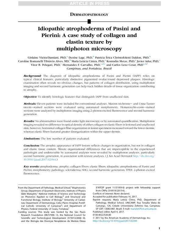
Background: The diagnosis of idiopathic atrophoderma of Pasini and Pierini (IAPP) relies on typical clinical features, particularly distinctive pigmented ovular/round depressed plaques. Histologic examination often reveals no obvious changes, but patterns of collagen distribution, using multiphoton imaging and second harmonic generation can help track hidden details of tissue organization contributing to atrophy. Objective: To identify histologic features that distinguish IAPP from unaffected skin. Methods: Eleven patients were included for conventional analyses. Masson trichrome– and Unna-Tanzer orcein–stained sections were evaluated using automated morphometry. Hematoxylin-eosin–stained sections were analyzed by multiphoton imaging using 2-photon excited fluorescence and second harmonic generation. Results: No abnormalities were found under light microscopy or by automated quantification. Multiphoton imaging revealed no difference in optical density of either collagen or elastic fibers in lesioned and unaffected skin; however, horizontal collagen fiber organization in lesion specimens increased toward the lower dermis, whereas elastic fibers featured greater disorganization within the upper dermis. Limitations: The low number of patients evaluated. Conclusion: The atrophic appearance of IAPP lesions reflects changes in organization, but not in collagen and elastic tissue content. Minute organizational differences that are imperceptible to the experienced pathologist and undetectable by automated analyses were revealed by multiphoton analyses, particularly second harmonic generation, in association with texture analyses.
Archivos asociados
Licencia
Identificadores
Colecciones
Articulos(SEDE CENTRAL)
Articulos de SEDE CENTRAL
Articulos de SEDE CENTRAL
Citación
Vieira Damiani, Gislaine; Lage, Denise; Daldon, Patrícia Érica Christofoletti ; Alves, Caroline Romanelli Tibúrcio ; Cintra, Maria Letícia; et al.; Idiopathic atrophoderma of Pasini and Pierini: A case study of collagen and elastin texture by multiphoton microscopy; Mosby-Elsevier; Journal of the American Academy of Dermatology; 77; 5; 11-2017; 930–937
Compartir
Altmétricas


