Mostrar el registro sencillo del ítem
dc.contributor.author
Szpilbarg, Natalia

dc.contributor.author
Castro-Parodi, Mauricio Omar

dc.contributor.author
Reppetti, Julieta

dc.contributor.author
Repetto, M.
dc.contributor.author
Maskin, B.
dc.contributor.author
Martinez, Nora Alicia

dc.contributor.author
Damiano, Alicia Ermelinda

dc.date.available
2018-03-14T18:06:12Z
dc.date.issued
2015-05
dc.identifier.citation
Szpilbarg, Natalia; Castro-Parodi, Mauricio Omar; Reppetti, Julieta; Repetto, M.; Maskin, B.; et al.; Placental programmed cell death: Insights into the role of aquaporins; Oxford University Press; Molecular Human Reproduction; 22; 1; 5-2015; 46-56
dc.identifier.issn
1360-9947
dc.identifier.uri
http://hdl.handle.net/11336/38757
dc.description.abstract
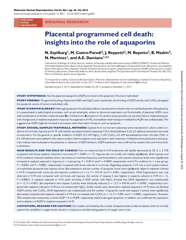
STUDY HYPOTHESIS: Are the placental aquaporins (AQPs) involved in the apoptosis of human trophoblast? STUDY FINDING: The general blocking of placental AQPs with HgCl2 and, in particular, the blocking of AQP3 activity with CuSO4 abrogated the apoptotic events of human trophoblast cells. WHAT IS KNOWN ALREADY: Although apoptosis of trophoblast cells is a natural event involved in the normal development of the placenta, it is exacerbated in pathological processes, such as pre-eclampsia, where an abnormal expression and functionality of placental AQPs occur without alterations in the feto-maternal water flux. Furthermore, fluctuations in O2 tension are proposed to be a potent inducer of placental apoptotic changes and, in explants exposed to hypoxia/reoxygenation (H/R), transcellular water transport mediated by AQPs was undetectable. This suggests that AQPs might be involved in processes other than water transport, such as apoptosis. STUDY DESIGN, SAMPLES/MATERIALS, METHODS: Explants from normal term placentas were maintained in culture under conditions of normoxia, hypoxia and H/R. Cell viability was determined by assessing 3-(4,5-dimethylthiazol-2-yl)-2,5-diphenyl tetrazolium bromide incorporation. For the general or specific inhibition of AQPs, 0.3 mM HgCl2, 5 mM CuSO4, 0.3 mM tetraethylammonium chloride (TEA) or 0.5 mM phloretin were added to the culture medium before explants were exposed to each treatment. Oxidative stress parameters and apoptotic indexes were evaluated in the presence or absence of AQPs blockers. AQP3 expression was confirmed by western blot and immunohistochemistry. MAIN RESULTS AND THE ROLE OF CHANCE: First, we observed that in H/R treatments cell viability decreased by 20.16 ± 5.73% compared with those explants cultured in normoxia (P = 0.009; n = 7). Hypoxia did not modify cell viability significantly. Both hypoxia and H/R conditions induced oxidative stress. Spontaneous chemiluminescence and thiobarbituric acid reactive substance levels were significantly increased in explants exposed to hypoxia (n = 6 per group, P = 0.0316 and P = 0.0009, respectively) and H/R conditions (n = 6 per group, P = 0.0281 and P = 0.0001, respectively) compared with those cultured in normoxia. Regarding apoptosis, H/R was a more potent inducer of trophoblast apoptosis than hypoxia alone. Bax expression and the number of apoptotic nuclei were significantly higher in explants cultured in H/R compared with normoxia and hypoxia conditions (n = 12, P = 0.0135 and P = 0.001, respectively). DNA fragmentation was only observed in H/R and, compared with normoxia and hypoxia, the activity of caspase-3 was highest in explants cultured in H/R (n = 12, P = 0.0001). In explants exposed to H/R, steric blocking of AQP activity with HgCl2 showed that DNA degradation was undetectable (n = 12, P = 0.001). Bax expression and caspase-3 activity were drastically reduced (n = 12, P = 0.0146 and P = 0.0001, respectively) compared with explants cultured in H/R but not treated with HgCl2. Similar results were observed in explants exposed to H/R when we blocked AQP3 activity with CuSO4. DNA degradation was undetectable and the number of apoptotic nuclei and caspase-3 activity were significantly decreased compared with explants cultured in H/R but not treated with CuSO4 (n = 12, P = 0.001 and P = 0.0001, respectively). However, TEA and phloretin treatments, to block AQP1/4 or AQP9, respectively, failed in abrogate apoptosis. In addition, we confirmed the expression and localization of AQP3 in explants exposed to H/R. LIMITATIONS, REASONS FOR CAUTION: Our studies are limited by the number of experimental conditions tested, which do not fully capture the variability in oxygen levels, duration of exposure and alternating patterns of oxygen seen in vivo. WIDER IMPLICATIONS OF THE FINDINGS: Our results suggest that any alteration in placental AQP expression might disturb the equilibrium of the normal apoptotic events and may be an underlying cause in the pathophysiology of placental gestational disorders such as pre-eclampsia. Furthermore, the dysregulation of placental AQPs may be one of the crucial factors in triggering the clinical manifestations of pre-eclampsia.
dc.format
application/pdf
dc.language.iso
eng
dc.publisher
Oxford University Press

dc.rights
info:eu-repo/semantics/openAccess
dc.rights.uri
https://creativecommons.org/licenses/by-nc-sa/2.5/ar/
dc.subject
Apoptosis
dc.subject
Aquaporins
dc.subject
Human Placenta
dc.subject
Intermittent Hypoxia
dc.subject
Pre-Eclampsia
dc.subject
Trophoblast
dc.subject.classification
Fisiología

dc.subject.classification
Medicina Básica

dc.subject.classification
CIENCIAS MÉDICAS Y DE LA SALUD

dc.title
Placental programmed cell death: Insights into the role of aquaporins
dc.type
info:eu-repo/semantics/article
dc.type
info:ar-repo/semantics/artículo
dc.type
info:eu-repo/semantics/publishedVersion
dc.date.updated
2018-03-14T17:13:42Z
dc.journal.volume
22
dc.journal.number
1
dc.journal.pagination
46-56
dc.journal.pais
Reino Unido

dc.journal.ciudad
Oxford
dc.description.fil
Fil: Szpilbarg, Natalia. Consejo Nacional de Investigaciones Científicas y Técnicas. Oficina de Coordinación Administrativa Houssay. Instituto de Fisiología y Biofísica Bernardo Houssay. Universidad de Buenos Aires. Facultad de Medicina. Instituto de Fisiología y Biofísica Bernardo Houssay; Argentina
dc.description.fil
Fil: Castro-Parodi, Mauricio Omar. Universidad de Buenos Aires. Facultad de Farmacia y Bioquímica. Departamento de Ciencias Biológicas; Argentina. Consejo Nacional de Investigaciones Científicas y Técnicas; Argentina
dc.description.fil
Fil: Reppetti, Julieta. Universidad de Buenos Aires. Facultad de Farmacia y Bioquímica. Departamento de Ciencias Biológicas; Argentina. Consejo Nacional de Investigaciones Científicas y Técnicas; Argentina
dc.description.fil
Fil: Repetto, M.. Universidad de Buenos Aires. Facultad de Farmacia y Bioquímica. Departamento de Química Analítica y Fisicoquímica. Cátedra de Química General e Inorgánica; Argentina
dc.description.fil
Fil: Maskin, B.. Hospital Nacional "Prof. Dr. Alejandro Posadas"; Argentina
dc.description.fil
Fil: Martinez, Nora Alicia. Consejo Nacional de Investigaciones Científicas y Técnicas. Oficina de Coordinación Administrativa Houssay. Instituto de Fisiología y Biofísica Bernardo Houssay. Universidad de Buenos Aires. Facultad de Medicina. Instituto de Fisiología y Biofísica Bernardo Houssay; Argentina
dc.description.fil
Fil: Damiano, Alicia Ermelinda. Consejo Nacional de Investigaciones Científicas y Técnicas. Oficina de Coordinación Administrativa Houssay. Instituto de Fisiología y Biofísica Bernardo Houssay. Universidad de Buenos Aires. Facultad de Medicina. Instituto de Fisiología y Biofísica Bernardo Houssay; Argentina. Universidad de Buenos Aires. Facultad de Farmacia y Bioquímica. Departamento de Ciencias Biológicas; Argentina
dc.journal.title
Molecular Human Reproduction

dc.relation.alternativeid
info:eu-repo/semantics/altIdentifier/doi/http://dx.doi.org/10.1093/molehr/gav063
dc.relation.alternativeid
info:eu-repo/semantics/altIdentifier/url/https://academic.oup.com/molehr/article/22/1/46/2459799
Archivos asociados
