Mostrar el registro sencillo del ítem
dc.contributor.author
Carrascosa, Patricia M.
dc.contributor.author
Cury, Ricardo C.
dc.contributor.author
Deviggiano, Alejandro
dc.contributor.author
Capunay, Carlos
dc.contributor.author
Campisi, Roxana
dc.contributor.author
López de Munain, Marina
dc.contributor.author
Vallejos, Javier
dc.contributor.author
Tajer, Carlos
dc.contributor.author
Rodriguez Granillo, Gaston Alfredo
dc.date.available
2018-03-06T19:35:36Z
dc.date.issued
2015-05
dc.identifier.citation
Carrascosa, Patricia M.; Cury, Ricardo C.; Deviggiano, Alejandro; Capunay, Carlos; Campisi, Roxana; et al.; Comparison of myocardial perfusion evaluation with single versus dual-energy ct and effect of beam-hardening artifacts; Elsevier Science Inc; Academic Radiology; 22; 5; 5-2015; 591-599
dc.identifier.issn
1076-6332
dc.identifier.uri
http://hdl.handle.net/11336/38034
dc.description.abstract
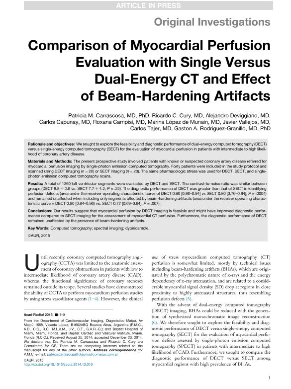
Rationale and objectives: We sought to explore the feasibility and diagnostic performance of dual-energy computed tomography (DECT) versus single-energy computed tomography (SECT) for the evaluation of myocardial perfusion in patients with intermediate to high likelihood of coronary artery disease. Materials and Methods: The present prospective study involved patients with known or suspected coronary artery disease referred for myocardial perfusion imaging by single-photon emission computed tomography. Forty patients were included in the study protocol and scanned using DECT imaging (n=20) or SECT imaging (n=20). The same pharmacologic stress was used for DECT, SECT, and single-photon emission computed tomography scans. Results: A total of 1360 left ventricular segments were evaluated by DECT and SECT. The contrast-to-noise ratio was similar between groups (DECT 8.8±2.9 vs. SECT 7.7±4.2; P = 22). The diagnostic performance of DECT was greater than that of SECT in identifying perfusion defects (area under the receiver operating characteristic curve of DECT 0.90 [0.86-0.94] vs SECT 0.80 [0.76-0.84]; P = 0004) and remained unaffected when including only segments affected by beam-hardening artifacts (area under the receiver operating characteristic curve=DECT 0.90 [0.84-0.96) vs. SECT 0.77 [0.69-0.84]; P = 007). Conclusions: Our results suggest that myocardial perfusion by DECT imaging is feasible and might have improved diagnostic performance compared to SECT imaging for the assessment of myocardial CT perfusion. Furthermore, the diagnostic performance of DECT remained unaffected by the presence of beam-hardening artifacts.
dc.format
application/pdf
dc.language.iso
eng
dc.publisher
Elsevier Science Inc
dc.rights
info:eu-repo/semantics/openAccess
dc.rights.uri
https://creativecommons.org/licenses/by-nc-nd/2.5/ar/
dc.subject
Computed Tomography
dc.subject
Dypiridamole
dc.subject
Spectral Imaging
dc.subject.classification
Medicina Critica y de Emergencia
dc.subject.classification
Medicina Clínica
dc.subject.classification
CIENCIAS MÉDICAS Y DE LA SALUD
dc.title
Comparison of myocardial perfusion evaluation with single versus dual-energy ct and effect of beam-hardening artifacts
dc.type
info:eu-repo/semantics/article
dc.type
info:ar-repo/semantics/artículo
dc.type
info:eu-repo/semantics/publishedVersion
dc.date.updated
2018-03-06T17:44:50Z
dc.journal.volume
22
dc.journal.number
5
dc.journal.pagination
591-599
dc.journal.pais
Países Bajos
dc.journal.ciudad
Amsterdam
dc.description.fil
Fil: Carrascosa, Patricia M.. Diagnóstico Maipú; Argentina
dc.description.fil
Fil: Cury, Ricardo C.. Baptist Cardiac and Vascular Institute; Estados Unidos. Baptist Hospital of Miami; Estados Unidos
dc.description.fil
Fil: Deviggiano, Alejandro. Diagnóstico Maipú; Argentina
dc.description.fil
Fil: Capunay, Carlos. Diagnóstico Maipú; Argentina
dc.description.fil
Fil: Campisi, Roxana. Diagnóstico Maipú; Argentina
dc.description.fil
Fil: López de Munain, Marina. Diagnóstico Maipú; Argentina
dc.description.fil
Fil: Vallejos, Javier. Diagnóstico Maipú; Argentina
dc.description.fil
Fil: Tajer, Carlos. Diagnóstico Maipú; Argentina
dc.description.fil
Fil: Rodriguez Granillo, Gaston Alfredo. Diagnóstico Maipú; Argentina. Consejo Nacional de Investigaciones Científicas y Técnicas; Argentina
dc.journal.title
Academic Radiology
dc.relation.alternativeid
info:eu-repo/semantics/altIdentifier/doi/http://dx.doi.org/10.1016/j.acra.2014.12.019
dc.relation.alternativeid
info:eu-repo/semantics/altIdentifier/url/https://www.sciencedirect.com/science/article/pii/S1076633215000094
Archivos asociados
Tamaño:
1.053Mb
Formato:
PDF