Artículo
American tegumentary leishmaniasis: T-cell differentiation profile of cutaneous and mucosal forms—co-infection with Trypanosoma cruzi
Parodi Ramoneda, Cecilia María ; Garcia Bustos, Maria Fernanda
 ; Barrio, Alejandra; Ramos, Federico
 ; González Prieto, Ana Gabriela
 ; Mora, Maria Celia
 ; Baré, Patricia
 ; Basombrío, Miguel Ángel Manuel
 ; de Elizalde, Maria Marta
 ; Garcia Bustos, Maria Fernanda
 ; Barrio, Alejandra; Ramos, Federico
 ; González Prieto, Ana Gabriela
 ; Mora, Maria Celia
 ; Baré, Patricia
 ; Basombrío, Miguel Ángel Manuel
 ; de Elizalde, Maria Marta
Fecha de publicación:
08/2016
Editorial:
Springer
Revista:
Medical Microbiology and Immunology
ISSN:
0300-8584
Idioma:
Inglés
Tipo de recurso:
Artículo publicado
Clasificación temática:
Resumen
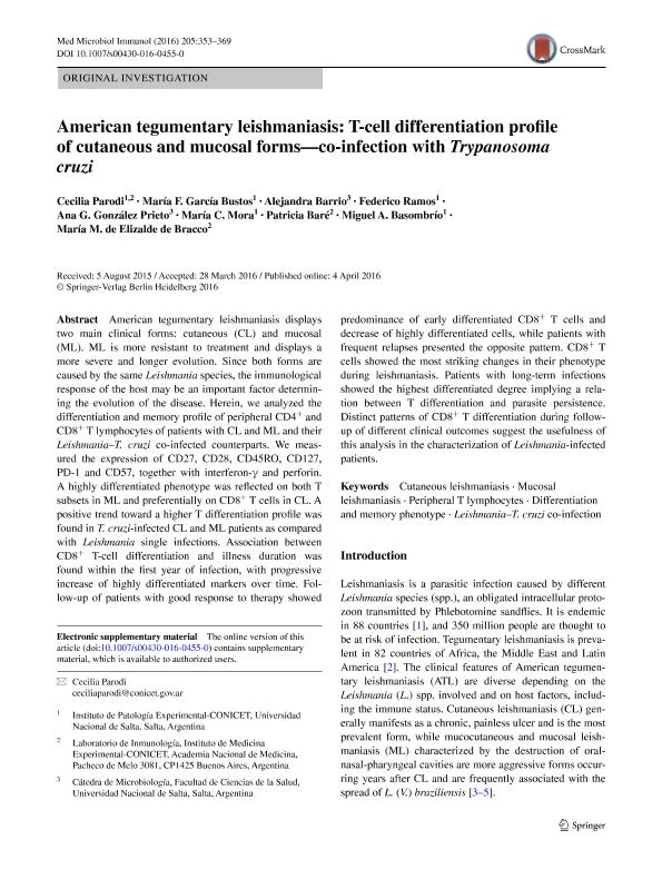
American tegumentary leishmaniasis displays two main clinical forms: cutaneous (CL) and mucosal (ML). ML is more resistant to treatment and displays a more severe and longer evolution. Since both forms are caused by the same Leishmania species, the immunological response of the host may be an important factor determining the evolution of the disease. Herein, we analyzed the differentiation and memory profile of peripheral CD4+ and CD8+ T lymphocytes of patients with CL and ML and their Leishmania–T. cruzi co-infected counterparts. We measured the expression of CD27, CD28, CD45RO, CD127, PD-1 and CD57, together with interferon-γ and perforin. A highly differentiated phenotype was reflected on both T subsets in ML and preferentially on CD8+ T cells in CL. A positive trend toward a higher T differentiation profile was found in T. cruzi-infected CL and ML patients as compared with Leishmania single infections. Association between CD8+ T-cell differentiation and illness duration was found within the first year of infection, with progressive increase of highly differentiated markers over time. Follow-up of patients with good response to therapy showed predominance of early differentiated CD8+ T cells and decrease of highly differentiated cells, while patients with frequent relapses presented the opposite pattern. CD8+ T cells showed the most striking changes in their phenotype during leishmaniasis. Patients with long-term infections showed the highest differentiated degree implying a relation between T differentiation and parasite persistence. Distinct patterns of CD8+ T differentiation during follow-up of different clinical outcomes suggest the usefulness of this analysis in the characterization of Leishmania-infected patients.
Archivos asociados
Licencia
Identificadores
Colecciones
Articulos(CCT - SALTA-JUJUY)
Articulos de CTRO.CIENTIFICO TECNOL.CONICET - SALTA-JUJUY
Articulos de CTRO.CIENTIFICO TECNOL.CONICET - SALTA-JUJUY
Articulos(IPE)
Articulos de INST.DE PATOLOGIA EXPERIMENTAL
Articulos de INST.DE PATOLOGIA EXPERIMENTAL
Citación
Parodi Ramoneda, Cecilia María; Garcia Bustos, Maria Fernanda; Barrio, Alejandra; Ramos, Federico; González Prieto, Ana Gabriela; et al.; American tegumentary leishmaniasis: T-cell differentiation profile of cutaneous and mucosal forms—co-infection with Trypanosoma cruzi; Springer; Medical Microbiology and Immunology; 205; 4; 8-2016; 353-369
Compartir
Altmétricas


