Mostrar el registro sencillo del ítem
dc.contributor.author
de Battista, Juan Carlos

dc.contributor.author
Andaluz, Norberto
dc.contributor.author
Zuccarello, Mario
dc.contributor.author
Keller, Jeffrey T.
dc.date.available
2018-01-23T17:24:21Z
dc.date.issued
2014-03
dc.identifier.citation
de Battista, Juan Carlos; Andaluz, Norberto; Zuccarello, Mario; Keller, Jeffrey T.; Refining the Indications for the Addition of Orbital Osteotomy during Anterior Cranial Base Approaches: Morphometric and Radiologic Study of the Anterior Cranial Base Osteology; Georg Thieme Verlag KG; Journal of neurological surgery reports; 75; 3-2014; 22-26
dc.identifier.issn
2193-6358
dc.identifier.uri
http://hdl.handle.net/11336/34268
dc.description.abstract
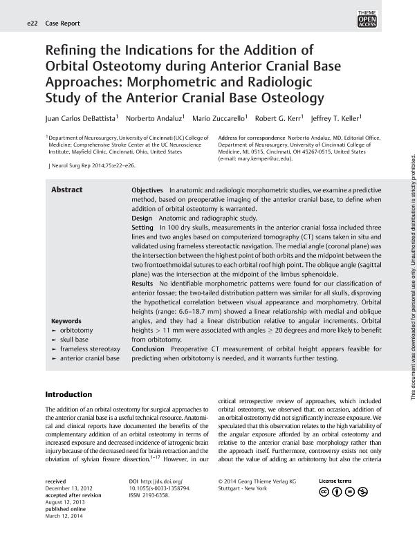
Objectives In anatomic and radiologic morphometric studies, we examine a predictive method, based on preoperative imaging of the anterior cranial base, to define when addition of orbital osteotomy is warranted. Design Anatomic and radiographic study. Setting In 100 dry skulls, measurements in the anterior cranial fossa included three lines and two angles based on computerized tomography (CT) scans taken in situ and validated using frameless stereotactic navigation. The medial angle (coronal plane) was the intersection between the highest point of both orbits and the midpoint between the two frontoethmoidal sutures to each orbital roof high point. The oblique angle (sagittal plane) was the intersection at the midpoint of the limbus sphenoidale. Results No identifiable morphometric patterns were found for our classification of anterior fossae; the two-tailed distribution pattern was similar for all skulls, disproving the hypothetical correlation between visual appearance and morphometry. Orbital heights (range: 6.6–18.7 mm) showed a linear relationship with medial and oblique angles, and they had a linear distribution relative to angular increments. Orbital heights > 11 mm were associated with angles ≥ 20 degrees and more likely to benefit from orbitotomy. Conclusion Preoperative CT measurement of orbital height appears feasible for predicting when orbitotomy is needed, and it warrants further testing.
dc.format
application/pdf
dc.language.iso
eng
dc.publisher
Georg Thieme Verlag KG
dc.rights
info:eu-repo/semantics/openAccess
dc.rights.uri
https://creativecommons.org/licenses/by-nc-sa/2.5/ar/
dc.subject
Orbitotomy
dc.subject
Skull Base
dc.subject
Frameless Stereotaxy
dc.subject
Anterior Cranial Base
dc.subject.classification
Inmunología

dc.subject.classification
Medicina Básica

dc.subject.classification
CIENCIAS MÉDICAS Y DE LA SALUD

dc.title
Refining the Indications for the Addition of Orbital Osteotomy during Anterior Cranial Base Approaches: Morphometric and Radiologic Study of the Anterior Cranial Base Osteology
dc.type
info:eu-repo/semantics/article
dc.type
info:ar-repo/semantics/artículo
dc.type
info:eu-repo/semantics/publishedVersion
dc.date.updated
2018-01-23T17:18:46Z
dc.journal.volume
75
dc.journal.pagination
22-26
dc.journal.pais
Estados Unidos

dc.journal.ciudad
New York
dc.description.fil
Fil: de Battista, Juan Carlos. Consejo Nacional de Investigaciones Científicas y Técnicas; Argentina. University of Cincinnati; Estados Unidos
dc.description.fil
Fil: Andaluz, Norberto. University of Cincinnati; Estados Unidos
dc.description.fil
Fil: Zuccarello, Mario. University of Cincinnati; Estados Unidos
dc.description.fil
Fil: Keller, Jeffrey T.. University of Cincinnati; Estados Unidos
dc.journal.title
Journal of neurological surgery reports
dc.relation.alternativeid
info:eu-repo/semantics/altIdentifier/doi/http://dx.doi.org/10.1055/s-0033-1358794
dc.relation.alternativeid
info:eu-repo/semantics/altIdentifier/url/https://www.thieme-connect.de/DOI/DOI?10.1055/s-0033-1358794
Archivos asociados
