Mostrar el registro sencillo del ítem
dc.contributor.author
Vanzulli, Silvia

dc.contributor.author
Efeyan, Alejo
dc.contributor.author
Benavides, Fernando
dc.contributor.author
Helguero, Luisa A.
dc.contributor.author
Peters, Giselle
dc.contributor.author
Shen, Jianjun
dc.contributor.author
Conti, Claudio J.
dc.contributor.author
Lanari, Claudia Lee Malvina

dc.contributor.author
Molinolo, Alfredo

dc.date.available
2017-12-22T00:48:16Z
dc.date.issued
2002-12
dc.identifier.citation
Molinolo, Alfredo; Lanari, Claudia Lee Malvina; Conti, Claudio J.; Shen, Jianjun; Peters, Giselle; Helguero, Luisa A.; et al.; p21,p27 and p53 in estrogen and antiprogestin- induced regression of experimental mouse mammary ductal carcinomas; Oxford University Press; Carcinogenesis; 23; 5; 12-2002; 749-757
dc.identifier.issn
0143-3334
dc.identifier.uri
http://hdl.handle.net/11336/31324
dc.description.abstract
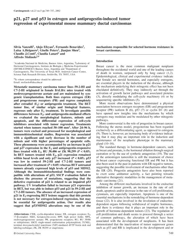
Metastatic mammary carcinoma tumor lines 59-2-HI and C7-2-HI originated in female BALB/c mice treated with medroxyprogesterone acetate and are maintained by syngeneic transplantation. Both lines express estrogen (ER) and progesterone receptors (PR) and regress completely after estradiol (E 2 ) or antiprogestin treatment. The BET tumor line, of similar origin and biological features, regresses only after E 2 treatment. To investigate possible differences between E 2 - and antiprogestin-mediated effects we evaluated the morphological features, mitosis and apoptosis, and the differential expression of cell-cycle inhibitors associated with tumor regression. Treatments started when tumors reached 50–100 mm 2 . After 24–96 h, tumors were excised and processed for morphological and immunohistochemical studies. Regression was associated with a significant and early decrease in the number of mitosis and with higher percentages of apoptotic cells. These phenomena were accompanied by an increase in p21 and p27 expression in the E 2 and antiprogestin-responsive lines treated with E 2 , RU 38.486 or ZK 98.299 ( P < 0.05). In BET tumors treated with E 2 , p21 expression remained within basal levels and only p27 increased ( P < 0.05). p53 was low in control 59-2-HI and C7-2-HI tumors and increased after treatment ( P < 0.05) whereas BET untreated tumors already expressed high levels of p53 and MDM2. Although the immunohistochemical findings were compatible with alterations of p53, SSCP evaluation failed to disclose the presence of mutations, suggesting that the defective expression of p21 is related to an impaired p53 pathway. UV irradiation failed to increase p21 expression in BET, but was able to induce p53 and p21 in 59-2-HI and C7-2-HI tumors. The absence of an increased expression of p21 in E 2 -regressing BET lesions suggests that this protein is not necessary for estrogen-induced regression, but may be essential for antiprogestin action. Our results also suggest that p53/MDM2 alterations may be one of the mechanisms responsible for selected hormone resistance in breast carcinomas.
dc.format
application/pdf
dc.language.iso
eng
dc.publisher
Oxford University Press

dc.rights
info:eu-repo/semantics/openAccess
dc.rights.uri
https://creativecommons.org/licenses/by-nc-sa/2.5/ar/
dc.subject
Apoptosis
dc.subject
Mammary Neoplasm
dc.subject
Tumor Supressor
dc.subject
Proto-Oncogene Proteins P21
dc.subject.classification
Bioquímica y Biología Molecular

dc.subject.classification
Medicina Básica

dc.subject.classification
CIENCIAS MÉDICAS Y DE LA SALUD

dc.subject.classification
Patología

dc.subject.classification
Medicina Básica

dc.subject.classification
CIENCIAS MÉDICAS Y DE LA SALUD

dc.title
p21,p27 and p53 in estrogen and antiprogestin- induced regression of experimental mouse mammary ductal carcinomas
dc.type
info:eu-repo/semantics/article
dc.type
info:ar-repo/semantics/artículo
dc.type
info:eu-repo/semantics/publishedVersion
dc.date.updated
2017-12-04T17:54:50Z
dc.identifier.eissn
1460-2180
dc.journal.volume
23
dc.journal.number
5
dc.journal.pagination
749-757
dc.journal.pais
Reino Unido

dc.journal.ciudad
Oxford
dc.description.fil
Fil: Vanzulli, Silvia. Academia Nacional de Medicina de Buenos Aires; Argentina
dc.description.fil
Fil: Efeyan, Alejo. Consejo Nacional de Investigaciones Científicas y Técnicas. Instituto de Biología y Medicina Experimental. Fundación de Instituto de Biología y Medicina Experimental. Instituto de Biología y Medicina Experimental; Argentina
dc.description.fil
Fil: Benavides, Fernando. University of Texas; Estados Unidos
dc.description.fil
Fil: Helguero, Luisa A.. Consejo Nacional de Investigaciones Científicas y Técnicas. Instituto de Biología y Medicina Experimental. Fundación de Instituto de Biología y Medicina Experimental. Instituto de Biología y Medicina Experimental; Argentina
dc.description.fil
Fil: Peters, Giselle. Consejo Nacional de Investigaciones Científicas y Técnicas. Instituto de Biología y Medicina Experimental. Fundación de Instituto de Biología y Medicina Experimental. Instituto de Biología y Medicina Experimental; Argentina
dc.description.fil
Fil: Shen, Jianjun. University of Texas; Estados Unidos
dc.description.fil
Fil: Conti, Claudio J.. University of Texas; Estados Unidos
dc.description.fil
Fil: Lanari, Claudia Lee Malvina. Consejo Nacional de Investigaciones Científicas y Técnicas. Instituto de Biología y Medicina Experimental. Fundación de Instituto de Biología y Medicina Experimental. Instituto de Biología y Medicina Experimental; Argentina
dc.description.fil
Fil: Molinolo, Alfredo. Consejo Nacional de Investigaciones Científicas y Técnicas. Instituto de Biología y Medicina Experimental. Fundación de Instituto de Biología y Medicina Experimental. Instituto de Biología y Medicina Experimental; Argentina
dc.journal.title
Carcinogenesis

dc.relation.alternativeid
info:eu-repo/semantics/altIdentifier/url/https://academic.oup.com/carcin/article/23/5/749/2608222
dc.relation.alternativeid
info:eu-repo/semantics/altIdentifier/doi/http://dx.doi.org/10.1093/carcin/23.5.749
dc.relation.alternativeid
info:eu-repo/semantics/altIdentifier/pmid/https://www.ncbi.nlm.nih.gov/pubmed/12016147
Archivos asociados
