Mostrar el registro sencillo del ítem
dc.contributor.author
Ireland, Alex

dc.contributor.author
Ferretti, Jose Luis

dc.contributor.author
Rittweger, J
dc.date.available
2017-12-20T18:22:51Z
dc.date.issued
2014-12
dc.identifier.citation
Ireland, Alex; Ferretti, Jose Luis; Rittweger, J; Imaging of the muscle-bone relationship; Springer; Current Osteoporosis Reports; 12; 4; 12-2014; 486-495
dc.identifier.issn
1544-2241
dc.identifier.uri
http://hdl.handle.net/11336/31134
dc.description.abstract
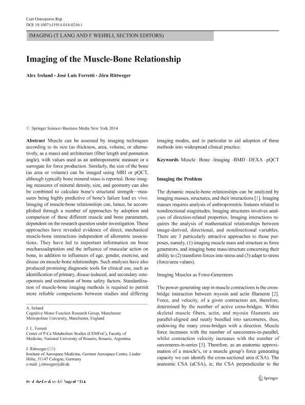
Muscle can be assessed by imaging techniques according to its size (as thickness, area, volume, or alternatively, as a mass) and architecture (fiber length and pennation angle), with values used as an anthropometric measure or a surrogate for force production. Similarly, the size of the bone (as area or volume) can be imaged using MRI or pQCT, although typically bone mineral mass is reported. Bone imaging measures of mineral density, size, and geometry can also be combined to calculate bone’s structural strength—measures being highly predictive of bone’s failure load ex vivo. Imaging of muscle-bone relationships can, hence, be accomplished through a number of approaches by adoption and comparison of these different muscle and bone parameters, dependent on the research question under investigation. These approaches have revealed evidence of direct, mechanical muscle-bone interactions independent of allometric associations. They have led to important information on bone mechanoadaptation and the influence of muscular action on bone, in addition to influences of age, gender, exercise, and disuse on muscle-bone relationships. Such analyses have also produced promising diagnostic tools for clinical use, such as identification of primary, disuse-induced, and secondary osteoporosis and estimation of bone safety factors. Standardization of muscle-bone imaging methods is required to permit more reliable comparisons between studies and differing imaging modes, and in particular to aid adoption of these methods into widespread clinical practice.
dc.format
application/pdf
dc.language.iso
eng
dc.publisher
Springer

dc.rights
info:eu-repo/semantics/openAccess
dc.rights.uri
https://creativecommons.org/licenses/by-nc-sa/2.5/ar/
dc.subject
Muscle
dc.subject
Bone
dc.subject
Imaging
dc.subject
Bmd
dc.subject
Dexa
dc.subject
Pqct
dc.subject.classification
Salud Ocupacional

dc.subject.classification
Ciencias de la Salud

dc.subject.classification
CIENCIAS MÉDICAS Y DE LA SALUD

dc.title
Imaging of the muscle-bone relationship
dc.type
info:eu-repo/semantics/article
dc.type
info:ar-repo/semantics/artículo
dc.type
info:eu-repo/semantics/publishedVersion
dc.date.updated
2017-12-12T18:20:34Z
dc.journal.volume
12
dc.journal.number
4
dc.journal.pagination
486-495
dc.journal.pais
Estados Unidos

dc.description.fil
Fil: Ireland, Alex. University of Manchester; Reino Unido
dc.description.fil
Fil: Ferretti, Jose Luis. Universidad Nacional de Rosario. Facultad de Ciencias Médicas. Centro de Estudios de Metabolismo Fosfocálcico; Argentina. Consejo Nacional de Investigaciones Científicas y Técnicas. Centro Científico Tecnológico Conicet - Rosario; Argentina
dc.description.fil
Fil: Rittweger, J. German Aerospace Centre; Alemania
dc.journal.title
Current Osteoporosis Reports
dc.relation.alternativeid
info:eu-repo/semantics/altIdentifier/doi/http://dx.doi.org/10.1007/s11914-014-0216-1
dc.relation.alternativeid
info:eu-repo/semantics/altIdentifier/url/https://link.springer.com/article/10.1007%2Fs11914-014-0216-1
Archivos asociados
