Artículo
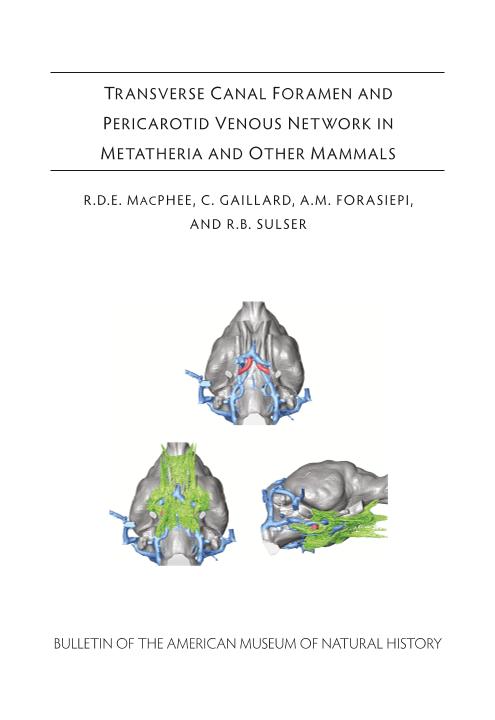
Transverse canal foramen and pericarotid venous network in metatheria and other mammals
Fecha de publicación:
06/2023
Editorial:
American Museum of Natural History
Revista:
Bulletin of the American Museum of Natural History
ISSN:
0003-0090
Idioma:
Inglés
Tipo de recurso:
Artículo publicado
Clasificación temática:
Resumen
Although few nondental features of the osteocranium consistently discriminate marsupials from placentals, the transverse canal foramen (TCF) has been repeatedly offered as a potential synapomorphy of crown-group Marsupialia and their closest allies. To explore this contention appropriately, the TCF needs to be evaluated in relation to the morphofunctional complex of which it is a part, something never previously undertaken in a systematic fashion. This complex, here defined as the pericarotid venous network (PCVN), is assessed using osteological, histological, and ontogenetic information. Although the TCF is usually thought of as a marsupial attribute, some living placentals also express it. What do these clades actually share in regard to this feature, and how do they differ? Our leading hypothesis is that the chief components of the PCVN begin development in the same way in both Marsupialia and Placentalia, but they follow different ontogenetic trajectories in terms of persistence, size, and connections with other elements of the cephalic venous vasculature. Similarities include shared presence of specific emissary and emissarylike veins in the mesocranial region that connect part of the endocranial dural vasculature (cavernous sinus or CS) to the systemic circulation (external and internal jugular veins plus the cerebrospinal venous system). In marsupials the principal pericarotid vessels are the transverse canal vein (TCV) and internal carotid vein (ICV). These veins almost always attain relatively large size during marsupial ontogeny. By contrast, in most placentals their apparent homologs (among others, emissary vein of the sphenoidal foramen and internal carotid venous plexus) evidently slow down or terminate their growth relatively early, and for this reason they play only a proportionally minor role in cephalic drainage in later life. In both clades, these vessels (informally grouped with others in the same region as pericarotid mesocranial distributaries, or PMDs) play a variable role in draining the CS in conjunction with the much larger petrosal sinuses. A pneumatic space within the basisphenoid - called the sphenoid sinus in placentals, transverse basisphenoid sinus (TBS) in marsupials - communicates with PCVN vasculature and should be considered an integral part of the network. The TBS contains red marrow tissues that are active centers of extramedullary hematopoiesis in young stages of some species, although how widespread this function may be in marsupial clades is not yet known. Previous explorations of the marsupial PCVN have been largely limited to determining whether, in any given taxon, a continuous passageway linking the right and left TCFs could be demonstrated running through the basisphenoid ("intramural" condition). It has long been known that a number of species apparently lack this particular passageway, and that the TCFs instead open into the braincase ("endocranial" condition). Puzzlingly, some species appear to have both passageways, others one or the other, and a few none at all, thus inviting questions about their equivalency and the circumstances under which the CS is actually drained by the TCV. Morphologically, these uncertainties can be resolved by viewing the full TCV as a tripartite entity, consisting of a trunk and rostral and caudal branches. The trunk, or the part that leaves the TCF for the external jugular system, receives the rostral and caudal branches, if both are present, within the body of the basisphenoid. The rostral or intramural branch has little or no direct communication with the endocranium in most investigated species. By contrast, the caudal or endocranial branch is an ordinary emissarium, in that it connects a part of the endocranial system of dural veins with the extracranial circulation. Determining branch routing alone does not adequately capture the scale of morphological variety and function encountered in marsupial PCVN organization. We distinguish five patterns of association between TCVs and other PCVN components. These patterns, based on both histological and osteological criteria, are defined as follows: (1) Simple: only rostral passageway present, caudal passageway absent or reduced to a thread; rostral branch veins form midline confluence within TBS in advance of hypophysis; minimal interaction with CS and its distributaries; rostral and caudal portions of TBS discontinuous. (2) Complex: mostly as in (1), except both rostral and caudal branches present and functional; caudal branches communicate with CS/ICV and do not form a confluence; TBS more extensive. (3) Compound: mostly as in (2), except TBS greatly expanded, incorporating most of rostral branch canals, which are correspondingly short. (4) Hybrid: differs from others in that only the pathways for enlarged caudal branches are significant; they originate from the CS/ICV caudal to the position of the hypophysis; rostral branches absent or highly reduced. (5) Indeterminate: transverse foramina, canals, and branches absent or unidentifiable as such, presumably due to vascular involution early in ontogeny. In light of TCV composition, the trunk of the TCV can be considered a mixed-origin vein, maximally receiving both a quasisystemic or emissarylike vessel (rostral branch) that does not originate from endocranial dural vessels, and a true emissarial vessel (caudal branch) that does. Some extant geomyoid rodents and strepsirrhine primates exhibit enlarged venous structures in the mesocranial region; these are briefly surveyed for comparative purposes, but resemblances to conditions in marsupials are superficial and unmistakably interpretable as convergences. Members of the extinct marsupial sister group Sparassodonta sometimes lack detectable TCFs, as do other non-marsupial metatherians in the fossil record. Evidence for the transverse canal and other PCVN components in other therians is briefly outlined. In summary, the development of mesocranial vasculature as outlined in this paper is hypothesized to be basal for therians, but Marsupialia and Placentalia radically differ in the end expression of PMDs in the adult stage. In prenatal stages of both clades, initial differentiation of these distributaries is presumably similar, but, compared to marsupials, in almost all placental groups these vessels are retained in an undeveloped or neotenic state. By contrast, enhanced expression of the TCV trunk and its branches seems to be a genuine novelty characterizing Marsupialia, although one probably present in some other metatherian groups. Accordingly, the transverse foramen, canal, and related features are probably best regarded as an innovation occurring in the marsupial stem, not a synapomorphy of the crown group as previously suggested by some authors.
Palabras clave:
Metatheria
,
Vasculature
,
Basicranium
Archivos asociados
Licencia
Identificadores
Colecciones
Articulos(IANIGLA)
Articulos de INST. ARG. DE NIVOLOGIA, GLACIOLOGIA Y CS. AMBIENT
Articulos de INST. ARG. DE NIVOLOGIA, GLACIOLOGIA Y CS. AMBIENT
Citación
Macphee, Ross Douglas Earle; Gaillard, Charlene; Forasiepi, Analia Marta; Sulser, R. Benjamin; Transverse canal foramen and pericarotid venous network in metatheria and other mammals; American Museum of Natural History; Bulletin of the American Museum of Natural History; 462; 1; 6-2023; 1-122
Compartir
Altmétricas


