Mostrar el registro sencillo del ítem
dc.contributor.author
Vangravs, Reinis
dc.contributor.author
Mezmale, Linda
dc.contributor.author
Slefarska Wolak, Daria
dc.contributor.author
Dauss, Edgars
dc.contributor.author
Ager, Clemens
dc.contributor.author
Corvalan, Alejandro H.
dc.contributor.author
Fernandez, Elmer Andres

dc.contributor.author
Mayhew, Chris A.
dc.contributor.author
Leja, Marcis
dc.contributor.author
Mochalski, Pawel
dc.date.available
2024-03-26T14:51:54Z
dc.date.issued
2024-03
dc.identifier.citation
Vangravs, Reinis; Mezmale, Linda; Slefarska Wolak, Daria; Dauss, Edgars; Ager, Clemens; et al.; Volatilomic signatures of different strains of Helicobacter pylori; Wiley Blackwell Publishing, Inc; Helicobacter; 29; 2; 3-2024; 1-11
dc.identifier.issn
1083-4389
dc.identifier.uri
http://hdl.handle.net/11336/231586
dc.description.abstract
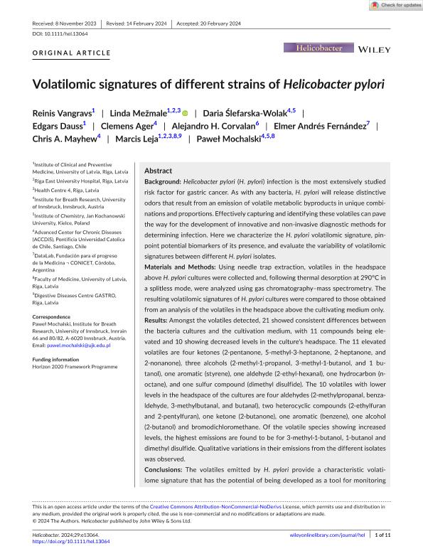
Background: Helicobacter pylori (H. pylori) infection is the most extensively studied risk factor for gastric cancer. As with any bacteria, H. pylori will release distinctive odors that result from an emission of volatile metabolic byproducts in unique combinations and proportions. Effectively capturing and identifying these volatiles can pave the way for the development of innovative and non-invasive diagnostic methods for determining infection. Here we characterize the H. pylori volatilomic signature, pinpoint potential biomarkers of its presence, and evaluate the variability of volatilomic signatures between different H. pylori isolates. Materials and Methods: Using needle trap extraction, volatiles in the headspace above H. pylori cultures were collected and, following thermal desorption at 290°C in a splitless mode, were analyzed using gas chromatography–mass spectrometry. The resulting volatilomic signatures of H. pylori cultures were compared to those obtained from an analysis of the volatiles in the headspace above the cultivating medium only. Results: Amongst the volatiles detected, 21 showed consistent differences between the bacteria cultures and the cultivation medium, with 11 compounds being elevated and 10 showing decreased levels in the culture's headspace. The 11 elevated volatiles are four ketones (2-pentanone, 5-methyl-3-heptanone, 2-heptanone, and 2-nonanone), three alcohols (2-methyl-1-propanol, 3-methyl-1-butanol, and 1 butanol), one aromatic (styrene), one aldehyde (2-ethyl-hexanal), one hydrocarbon (noctane), and one sulfur compound (dimethyl disulfide). The 10 volatiles with lower levels in the headspace of the cultures are four aldehydes (2-methylpropanal, benzaldehyde, 3-methylbutanal, and butanal), two heterocyclic compounds (2-ethylfuran and 2-pentylfuran), one ketone (2-butanone), one aromatic (benzene), one alcohol (2-butanol) and bromodichloromethane. Of the volatile species showing increased levels, the highest emissions are found to be for 3-methyl-1-butanol, 1-butanol and dimethyl disulfide. Qualitative variations in their emissions from the different isolates was observed. Conclusions: The volatiles emitted by H. pylori provide a characteristic volatilome signature that has the potential of being developed as a tool for monitoring infections caused by this pathogen. Furthermore, using the volatilome signature, we are able to differentiate different isolates of H. pylori. However, the volatiles also represent potential confounders for the recognition of gastric cancer volatile markers.
dc.format
application/pdf
dc.language.iso
eng
dc.publisher
Wiley Blackwell Publishing, Inc

dc.rights
info:eu-repo/semantics/openAccess
dc.rights.uri
https://creativecommons.org/licenses/by-nc-nd/2.5/ar/
dc.subject
cancer
dc.subject
gastrico
dc.subject
helicobacter
dc.subject.classification
Bioquímica y Biología Molecular

dc.subject.classification
Ciencias Biológicas

dc.subject.classification
CIENCIAS NATURALES Y EXACTAS

dc.title
Volatilomic signatures of different strains of Helicobacter pylori
dc.type
info:eu-repo/semantics/article
dc.type
info:ar-repo/semantics/artículo
dc.type
info:eu-repo/semantics/publishedVersion
dc.date.updated
2024-03-26T13:31:56Z
dc.identifier.eissn
1523-5378
dc.journal.volume
29
dc.journal.number
2
dc.journal.pagination
1-11
dc.journal.pais
Reino Unido

dc.description.fil
Fil: Vangravs, Reinis. Universidad de Letonia; Letonia
dc.description.fil
Fil: Mezmale, Linda. Universidad de Letonia; Letonia
dc.description.fil
Fil: Slefarska Wolak, Daria. University of Innsbruck; Austria
dc.description.fil
Fil: Dauss, Edgars. Universidad de Letonia; Letonia
dc.description.fil
Fil: Ager, Clemens. Universidad de Letonia; Letonia
dc.description.fil
Fil: Corvalan, Alejandro H.. Pontificia Universidad Católica de Chile; Chile
dc.description.fil
Fil: Fernandez, Elmer Andres. Consejo Nacional de Investigaciones Científicas y Técnicas. Centro Científico Tecnológico Conicet - Córdoba; Argentina. Fundación para el Progreso de la Medicina; Argentina
dc.description.fil
Fil: Mayhew, Chris A.. Universidad de Innsbruck; Austria
dc.description.fil
Fil: Leja, Marcis. Universidad de Letonia; Letonia
dc.description.fil
Fil: Mochalski, Pawel. Universidad de Innsbruck; Austria
dc.journal.title
Helicobacter

dc.relation.alternativeid
info:eu-repo/semantics/altIdentifier/url/https://onlinelibrary.wiley.com/doi/10.1111/hel.13064
dc.relation.alternativeid
info:eu-repo/semantics/altIdentifier/doi/http://dx.doi.org/10.1111/hel.13064
Archivos asociados
