Mostrar el registro sencillo del ítem
dc.contributor.author
Cruz Flores, Cesar

dc.contributor.author
Rodríguez, Cristian

dc.contributor.author
Giai, Constanza

dc.contributor.author
Vega, Israel Aníbal

dc.contributor.author
Castro Vazquez, Alfredo Juan

dc.date.available
2024-03-14T11:51:21Z
dc.date.issued
2023-10
dc.identifier.citation
Cruz Flores, Cesar; Rodríguez, Cristian; Giai, Constanza; Vega, Israel Aníbal; Castro Vazquez, Alfredo Juan; Pathogenesis of an experimental mycobacteriosis in an apple snail; Frontiers Media; Frontiers in Immunology; 14; 10-2023; 1-13
dc.identifier.issn
1664-3224
dc.identifier.uri
http://hdl.handle.net/11336/230502
dc.description.abstract
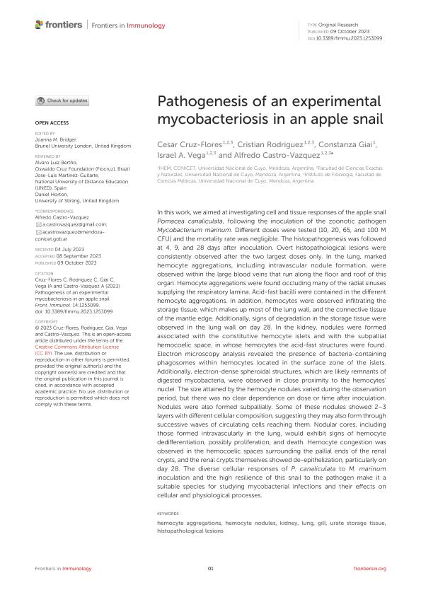
In this work, we aimed at investigating cell and tissue responses of the apple snail Pomacea canaliculata, following the inoculation of the zoonotic pathogen Mycobacterium marinum. Different doses were tested (10, 20, 65, and 100 M CFU) and the mortality rate was negligible. The histopathogenesis was followed at 4, 9, and 28 days after inoculation. Overt histopathological lesions were consistently observed after the two largest doses only. In the lung, marked hemocyte aggregations, including intravascular nodule formation, were observed within the large blood veins that run along the floor and roof of this organ. Hemocyte aggregations were found occluding many of the radial sinuses supplying the respiratory lamina. Acid-fast bacilli were contained in the different hemocyte aggregations. In addition, hemocytes were observed infiltrating the storage tissue, which makes up most of the lung wall, and the connective tissue of the mantle edge. Additionally, signs of degradation in the storage tissue were observed in the lung wall on day 28. In the kidney, nodules were formed associated with the constitutive hemocyte islets and with the subpallial hemocoelic space, in whose hemocytes the acid-fast structures were found. Electron microscopy analysis revealed the presence of bacteria-containing phagosomes within hemocytes located in the surface zone of the islets. Additionally, electron-dense spheroidal structures, which are likely remnants of digested mycobacteria, were observed in close proximity to the hemocytes’ nuclei. The size attained by the hemocyte nodules varied during the observation period, but there was no clear dependence on dose or time after inoculation. Nodules were also formed subpallially. Some of these nodules showed 2–3 layers with different cellular composition, suggesting they may also form through successive waves of circulating cells reaching them. Nodular cores, including those formed intravascularly in the lung, would exhibit signs of hemocyte dedifferentiation, possibly proliferation, and death. Hemocyte congestion was observed in the hemocoelic spaces surrounding the pallial ends of the renal crypts, and the renal crypts themselves showed de-epithelization, particularly on day 28. The diverse cellular responses of P. canaliculata to M. marinum inoculation and the high resilience of this snail to the pathogen make it a suitable species for studying mycobacterial infections and their effects on cellular and physiological processes.
dc.format
application/pdf
dc.language.iso
eng
dc.publisher
Frontiers Media

dc.rights
info:eu-repo/semantics/openAccess
dc.rights.uri
https://creativecommons.org/licenses/by-nc-sa/2.5/ar/
dc.subject
HEMOCYTE AGGREGATIONS
dc.subject
HEMOCYTE NODULES
dc.subject
KIDNEY
dc.subject
LUNG
dc.subject
GILL
dc.subject
URATE STORAGE TISSUE
dc.subject
HISTOPATOLOGICAL LESIONS
dc.subject.classification
Biología Celular, Microbiología

dc.subject.classification
Ciencias Biológicas

dc.subject.classification
CIENCIAS NATURALES Y EXACTAS

dc.title
Pathogenesis of an experimental mycobacteriosis in an apple snail
dc.type
info:eu-repo/semantics/article
dc.type
info:ar-repo/semantics/artículo
dc.type
info:eu-repo/semantics/publishedVersion
dc.date.updated
2024-03-13T14:23:58Z
dc.journal.volume
14
dc.journal.pagination
1-13
dc.journal.pais
Suiza

dc.description.fil
Fil: Cruz Flores, Cesar. Consejo Nacional de Investigaciones Científicas y Técnicas. Centro Científico Tecnológico Conicet - Mendoza. Instituto de Histología y Embriología de Mendoza Dr. Mario H. Burgos. Universidad Nacional de Cuyo. Facultad de Ciencias Médicas. Instituto de Histología y Embriología de Mendoza Dr. Mario H. Burgos; Argentina. Universidad Nacional de Cuyo. Facultad de Ciencias Exactas y Naturales. Departamento de Biología; Argentina
dc.description.fil
Fil: Rodríguez, Cristian. Universidad Nacional de Cuyo. Facultad de Ciencias Exactas y Naturales. Departamento de Biología; Argentina. Consejo Nacional de Investigaciones Científicas y Técnicas. Centro Científico Tecnológico Conicet - Mendoza. Instituto de Histología y Embriología de Mendoza Dr. Mario H. Burgos. Universidad Nacional de Cuyo. Facultad de Ciencias Médicas. Instituto de Histología y Embriología de Mendoza Dr. Mario H. Burgos; Argentina
dc.description.fil
Fil: Giai, Constanza. Consejo Nacional de Investigaciones Científicas y Técnicas. Centro Científico Tecnológico Conicet - Mendoza. Instituto de Histología y Embriología de Mendoza Dr. Mario H. Burgos. Universidad Nacional de Cuyo. Facultad de Ciencias Médicas. Instituto de Histología y Embriología de Mendoza Dr. Mario H. Burgos; Argentina
dc.description.fil
Fil: Vega, Israel Aníbal. Universidad Nacional de Cuyo. Facultad de Ciencias Exactas y Naturales. Departamento de Biología; Argentina. Universidad del Aconcagua. Facultad de Ciencias Médicas; Argentina. Consejo Nacional de Investigaciones Científicas y Técnicas. Centro Científico Tecnológico Conicet - Mendoza. Instituto de Histología y Embriología de Mendoza Dr. Mario H. Burgos. Universidad Nacional de Cuyo. Facultad de Ciencias Médicas. Instituto de Histología y Embriología de Mendoza Dr. Mario H. Burgos; Argentina
dc.description.fil
Fil: Castro Vazquez, Alfredo Juan. Consejo Nacional de Investigaciones Científicas y Técnicas. Centro Científico Tecnológico Conicet - Mendoza. Instituto de Histología y Embriología de Mendoza Dr. Mario H. Burgos. Universidad Nacional de Cuyo. Facultad de Ciencias Médicas. Instituto de Histología y Embriología de Mendoza Dr. Mario H. Burgos; Argentina. Universidad Nacional de Cuyo. Facultad de Ciencias Exactas y Naturales. Departamento de Biología; Argentina
dc.journal.title
Frontiers in Immunology
dc.relation.alternativeid
info:eu-repo/semantics/altIdentifier/url/https://www.frontiersin.org/articles/10.3389/fimmu.2023.1253099/full
dc.relation.alternativeid
info:eu-repo/semantics/altIdentifier/doi/http://dx.doi.org/10.3389/fimmu.2023.1253099
Archivos asociados
