Artículo
Promoting early neovascularization by allotransplanted adipose-derived Muse cells in an ovine model of acute myocardial infarction
Castillo Velasquez, Martha Giovanna ; Peralta, Tomás M.; Locatelli, Paola
 ; Velazquez, Candela
 ; Herrero, Yamila
 ; Crottogini, Alberto Jose
 ; Olea, Fernanda Daniela
 ; Cuniberti, Luis Alberto
 ; Peralta, Tomás M.; Locatelli, Paola
 ; Velazquez, Candela
 ; Herrero, Yamila
 ; Crottogini, Alberto Jose
 ; Olea, Fernanda Daniela
 ; Cuniberti, Luis Alberto
Fecha de publicación:
01/2023
Editorial:
Public Library of Science
Revista:
Plos One
ISSN:
1932-6203
Idioma:
Inglés
Tipo de recurso:
Artículo publicado
Clasificación temática:
Resumen
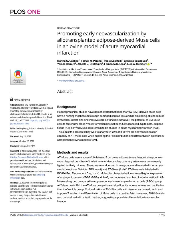
Background Recent preclinical studies have demonstrated that bone marrow (BM)-derived Muse cells have a homing mechanism to reach damaged cardiac tissue while also being able to reduce myocardial infarct size and improve cardiac function; however, the potential of BM-Muse cells to foster new blood-vessel formation has not been fully assessed. Up to date, adipose tissue (AT)-derived Muse cells remain to be studied in acute myocardial infarction (AMI). The aim of the present study was to analyze in vitro and in vivo the neovascularization capacity of AT-Muse cells while exploring their biodistribution and differentiation potential in a translational ovine model of AMI. Methods and results AT-Muse cells were successfully isolated from ovine adipose tissue. In adult sheep, one or more diagonal branches of the left anterior descending coronary artery were permanently ligated for thirty minutes. Sheep were randomized in two groups and treated with intramyocardial injections: Vehicle (PBS, n = 4) and AT-Muse (2x10⁷ AT-Muse cells labeled with PKH26 Red Fluorescent Dye, n = 4). Molecular characterization showed higher expression of angiogenic genes (VEGF, PGF and ANG) and increased number of tube formation in AT-Muse cells group compared to Adipose-derived mesenchymal stromal cells (ASCs) group. At 7 days post-IAM, the AT-Muse group showed significantly more arterioles and capillaries than the Vehicle group. Co-localization of PKH26+ cells with desmin, sarcomeric actin and troponin T implied the differentiation of Muse cells to a cardiac fate; moreover, PKH26+ cells also co-localized with a lectin marker, suggesting a possible differentiation to a vascular lineage. Conclusion Intramyocardially administered AT-Muse cells displayed a significant neovascularization activity and survival capacity in an ovine model of AMI.
Palabras clave:
Muse cells
,
ovine
,
myocardial infarction
Archivos asociados
Licencia
Identificadores
Colecciones
Articulos (IMETTYB)
Articulos de INSTITUTO DE MEDICINA TRASLACIONAL, TRASPLANTE Y BIOINGENIERIA
Articulos de INSTITUTO DE MEDICINA TRASLACIONAL, TRASPLANTE Y BIOINGENIERIA
Articulos(IBYME)
Articulos de INST.DE BIOLOGIA Y MEDICINA EXPERIMENTAL (I)
Articulos de INST.DE BIOLOGIA Y MEDICINA EXPERIMENTAL (I)
Citación
Castillo Velasquez, Martha Giovanna; Peralta, Tomás M.; Locatelli, Paola; Velazquez, Candela; Herrero, Yamila; et al.; Promoting early neovascularization by allotransplanted adipose-derived Muse cells in an ovine model of acute myocardial infarction; Public Library of Science; Plos One; 18; 1-2023; 1-15
Compartir
Altmétricas


