Mostrar el registro sencillo del ítem
dc.contributor.author
Holubiec, Mariana Ines

dc.contributor.author
Gellert, M.
dc.contributor.author
Hanschmann, E. M.
dc.date.available
2023-09-28T18:13:07Z
dc.date.issued
2022-11
dc.identifier.citation
Holubiec, Mariana Ines; Gellert, M.; Hanschmann, E. M.; Redox signaling and metabolism in Alzheimer's disease; Frontiers Media; Frontiers in Aging Neuroscience; 14; 11-2022; 1-17
dc.identifier.issn
1663-4365
dc.identifier.uri
http://hdl.handle.net/11336/213492
dc.description.abstract
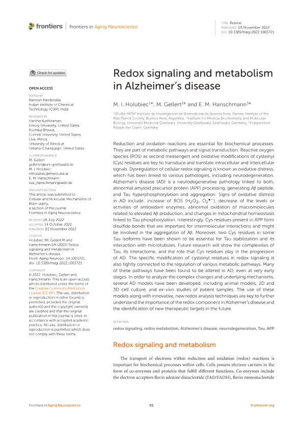
Reduction and oxidation reactions are essential for biochemical processes. They are part of metabolic pathways and signal transduction. Reactive oxygen species (ROS) as second messengers and oxidative modifications of cysteinyl (Cys) residues are key to transduce and translate intracellular and intercellular signals. Dysregulation of cellular redox signaling is known as oxidative distress, which has been linked to various pathologies, including neurodegeneration. Alzheimer's disease (AD) is a neurodegenerative pathology linked to both, abnormal amyloid precursor protein (APP) processing, generating Aβ peptide, and Tau hyperphosphorylation and aggregation. Signs of oxidative distress in AD include: increase of ROS (H2O2, O2•−), decrease of the levels or activities of antioxidant enzymes, abnormal oxidation of macromolecules related to elevated Aβ production, and changes in mitochondrial homeostasis linked to Tau phosphorylation. Interestingly, Cys residues present in APP form disulfide bonds that are important for intermolecular interactions and might be involved in the aggregation of Aβ. Moreover, two Cys residues in some Tau isoforms have been shown to be essential for Tau stabilization and its interaction with microtubules. Future research will show the complexities of Tau, its interactome, and the role that Cys residues play in the progression of AD. The specific modification of cysteinyl residues in redox signaling is also tightly connected to the regulation of various metabolic pathways. Many of these pathways have been found to be altered in AD, even at very early stages. In order to analyze the complex changes and underlying mechanisms, several AD models have been developed, including animal models, 2D and 3D cell culture, and ex-vivo studies of patient samples. The use of these models along with innovative, new redox analysis techniques are key to further understand the importance of the redox component in Alzheimer's disease and the identification of new therapeutic targets in the future.
dc.format
application/pdf
dc.language.iso
eng
dc.publisher
Frontiers Media

dc.rights
info:eu-repo/semantics/openAccess
dc.rights.uri
https://creativecommons.org/licenses/by/2.5/ar/
dc.subject
ALZHEIMER'S DISEASE
dc.subject
APP
dc.subject
NEURODEGENERATION
dc.subject
REDOX METABOLISM
dc.subject
REDOX SIGNALING
dc.subject
TAU
dc.subject.classification
Bioquímica y Biología Molecular

dc.subject.classification
Ciencias Biológicas

dc.subject.classification
CIENCIAS NATURALES Y EXACTAS

dc.title
Redox signaling and metabolism in Alzheimer's disease
dc.type
info:eu-repo/semantics/article
dc.type
info:ar-repo/semantics/artículo
dc.type
info:eu-repo/semantics/publishedVersion
dc.date.updated
2023-07-10T11:48:41Z
dc.journal.volume
14
dc.journal.pagination
1-17
dc.journal.pais
Suiza

dc.journal.ciudad
Lausana
dc.description.fil
Fil: Holubiec, Mariana Ines. Consejo Nacional de Investigaciones Científicas y Técnicas. Oficina de Coordinación Administrativa Parque Centenario. Instituto de Investigación en Biomedicina de Buenos Aires - Instituto Partner de la Sociedad Max Planck; Argentina
dc.description.fil
Fil: Gellert, M.. University Greifswald; Alemania
dc.description.fil
Fil: Hanschmann, E. M.. No especifíca;
dc.journal.title
Frontiers in Aging Neuroscience
dc.relation.alternativeid
info:eu-repo/semantics/altIdentifier/url/https://www.frontiersin.org/articles/10.3389/fnagi.2022.1003721/full
dc.relation.alternativeid
info:eu-repo/semantics/altIdentifier/doi/https://doi.org/10.3389/fnagi.2022.1003721
Archivos asociados
