Mostrar el registro sencillo del ítem
dc.contributor.author
Bonfili, Noelia Sabrina

dc.contributor.author
Barbeito Andrés, Jimena

dc.contributor.author
Bernal, Valeria

dc.contributor.author
Hallgrimsson, Benedikt

dc.contributor.author
Gonzalez, Paula Natalia

dc.date.available
2023-09-25T18:47:51Z
dc.date.issued
2022-03
dc.identifier.citation
Bonfili, Noelia Sabrina; Barbeito Andrés, Jimena; Bernal, Valeria; Hallgrimsson, Benedikt; Gonzalez, Paula Natalia; Morphological correspondence between brain and endocranial surfaces in mice exposed to undernutrition during development; Wiley Blackwell Publishing, Inc; Journal of Anatomy; 241; 1; 3-2022; 1-12
dc.identifier.issn
0021-8782
dc.identifier.uri
http://hdl.handle.net/11336/212970
dc.description.abstract
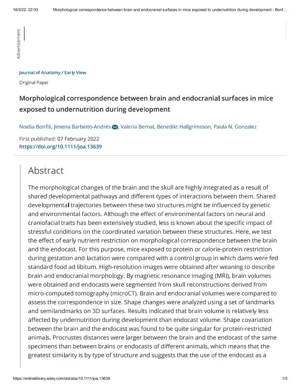
The morphological changes of the brain and the skull are highly integrated as a result of shared developmental pathways and different types of interactions between them. Shared developmental trajectories between these two structures might be influenced by genetic and environmental factors. Although the effect of environmental factors on neural and craniofacial traits has been extensively studied, less is known about the specific impact of stressful conditions on the coordinated variation between these structures. Here, we test the effect of early nutrient restriction on morphological correspondence between the brain and the endocast. For this purpose, mice exposed to protein or calorie-protein restriction during gestation and lactation were compared with a control group in which dams were fed standard food ad libitum. High-resolution images were obtained after weaning to describe brain and endocranial morphology. By magnetic resonance imaging (MRI), brain volumes were obtained and endocasts were segmented from skull reconstructions derived from micro-computed tomography (microCT). Brain and endocranial volumes were compared to assess the correspondence in size. Shape changes were analyzed using a set of landmarks and semilandmarks on 3D surfaces. Results indicated that brain volume is relatively less affected by undernutrition during development than endocast volume. Shape covariation between the brain and the endocast was found to be quite singular for protein-restricted animals. Procrustes distances were larger between the brain and the endocast of the same specimens than between brains or endocasts of different animals, which means that the greatest similarity is by type of structure and suggests that the use of the endocast as a direct proxy of the brain at this intraspecific scale could have some limitations. In the same line, patterns of brain shape asymmetry were not directly estimated from endocranial surfaces. In sum, our findings indicate that morphological variation and association between the brain and the endocast is modulated by environmental factors and support the idea that head morphogenesis results from complex processes that are sensitive to the pervasive influence of nutrient intake.
dc.format
application/pdf
dc.language.iso
eng
dc.publisher
Wiley Blackwell Publishing, Inc

dc.rights
info:eu-repo/semantics/openAccess
dc.rights.uri
https://creativecommons.org/licenses/by-nc-sa/2.5/ar/
dc.subject
BRAIN PLASTICITY
dc.subject
COMPUTED TOMOGRAPHY
dc.subject
GEOMETRIC MORPHOMETRICS
dc.subject
MAGNETIC RESONANCE IMAGING
dc.subject
MORPHOLOGICAL INTEGRATION
dc.subject
PALEONEUROLOGY
dc.subject.classification
Otros Tópicos Biológicos

dc.subject.classification
Ciencias Biológicas

dc.subject.classification
CIENCIAS NATURALES Y EXACTAS

dc.title
Morphological correspondence between brain and endocranial surfaces in mice exposed to undernutrition during development
dc.type
info:eu-repo/semantics/article
dc.type
info:ar-repo/semantics/artículo
dc.type
info:eu-repo/semantics/publishedVersion
dc.date.updated
2023-07-07T18:43:56Z
dc.journal.volume
241
dc.journal.number
1
dc.journal.pagination
1-12
dc.journal.pais
Reino Unido

dc.journal.ciudad
Londres
dc.description.fil
Fil: Bonfili, Noelia Sabrina. Universidad Nacional Arturo Jauretche; Argentina. Universidad Nacional Arturo Jauretche. Unidad Ejecutora de Estudios en Neurociencias y Sistemas Complejos. Provincia de Buenos Aires. Ministerio de Salud. Hospital Alta Complejidad en Red El Cruce Dr. Néstor Carlos Kirchner Samic. Unidad Ejecutora de Estudios en Neurociencias y Sistemas Complejos. Consejo Nacional de Investigaciones Científicas y Técnicas. Centro Científico Tecnológico Conicet - La Plata. Unidad Ejecutora de Estudios en Neurociencias y Sistemas Complejos; Argentina
dc.description.fil
Fil: Barbeito Andrés, Jimena. Universidad Nacional Arturo Jauretche; Argentina. Universidad Nacional Arturo Jauretche. Unidad Ejecutora de Estudios en Neurociencias y Sistemas Complejos. Provincia de Buenos Aires. Ministerio de Salud. Hospital Alta Complejidad en Red El Cruce Dr. Néstor Carlos Kirchner Samic. Unidad Ejecutora de Estudios en Neurociencias y Sistemas Complejos. Consejo Nacional de Investigaciones Científicas y Técnicas. Centro Científico Tecnológico Conicet - La Plata. Unidad Ejecutora de Estudios en Neurociencias y Sistemas Complejos; Argentina
dc.description.fil
Fil: Bernal, Valeria. Universidad Nacional de La Plata. Facultad de Ciencias Naturales y Museo. División Antropología; Argentina. Consejo Nacional de Investigaciones Científicas y Técnicas. Centro Científico Tecnológico Conicet - La Plata; Argentina
dc.description.fil
Fil: Hallgrimsson, Benedikt. University of Calgary; Canadá
dc.description.fil
Fil: Gonzalez, Paula Natalia. Universidad Nacional Arturo Jauretche; Argentina. Universidad Nacional Arturo Jauretche. Unidad Ejecutora de Estudios en Neurociencias y Sistemas Complejos. Provincia de Buenos Aires. Ministerio de Salud. Hospital Alta Complejidad en Red El Cruce Dr. Néstor Carlos Kirchner Samic. Unidad Ejecutora de Estudios en Neurociencias y Sistemas Complejos. Consejo Nacional de Investigaciones Científicas y Técnicas. Centro Científico Tecnológico Conicet - La Plata. Unidad Ejecutora de Estudios en Neurociencias y Sistemas Complejos; Argentina
dc.journal.title
Journal of Anatomy

dc.relation.alternativeid
info:eu-repo/semantics/altIdentifier/doi/http://dx.doi.org/10.1111/joa.13639
dc.relation.alternativeid
info:eu-repo/semantics/altIdentifier/url/https://onlinelibrary.wiley.com/doi/10.1111/joa.13639
Archivos asociados
