Mostrar el registro sencillo del ítem
dc.contributor.author
Fujisawa, Kyoko
dc.contributor.author
Silcott Niles, Charlotte
dc.contributor.author
Simonson, Poppy
dc.contributor.author
Lamattina, Daniela

dc.contributor.author
Humeres, Cristian Alejandro

dc.contributor.author
Bhattacharyya, Tapan

dc.contributor.author
Mertens, Pascal
dc.contributor.author
Thunissen, Caroline
dc.contributor.author
O'Rourke, Victoria
dc.contributor.author
Panczuk, Magdalena
dc.contributor.author
Whitworth, James A.
dc.contributor.author
Salomón, Oscar Daniel

dc.contributor.author
Miles, Michael A.

dc.date.available
2023-08-14T12:18:15Z
dc.date.issued
2021-07
dc.identifier.citation
Fujisawa, Kyoko; Silcott Niles, Charlotte; Simonson, Poppy; Lamattina, Daniela; Humeres, Cristian Alejandro; et al.; Emergent canine visceral leishmaniasis in Argentina: Comparative diagnostics and relevance to proliferation of human disease; Public Library of Science; Neglected Tropical Diseases; 15; 7; 7-2021; 1-19
dc.identifier.issn
1935-2735
dc.identifier.uri
http://hdl.handle.net/11336/208078
dc.description.abstract
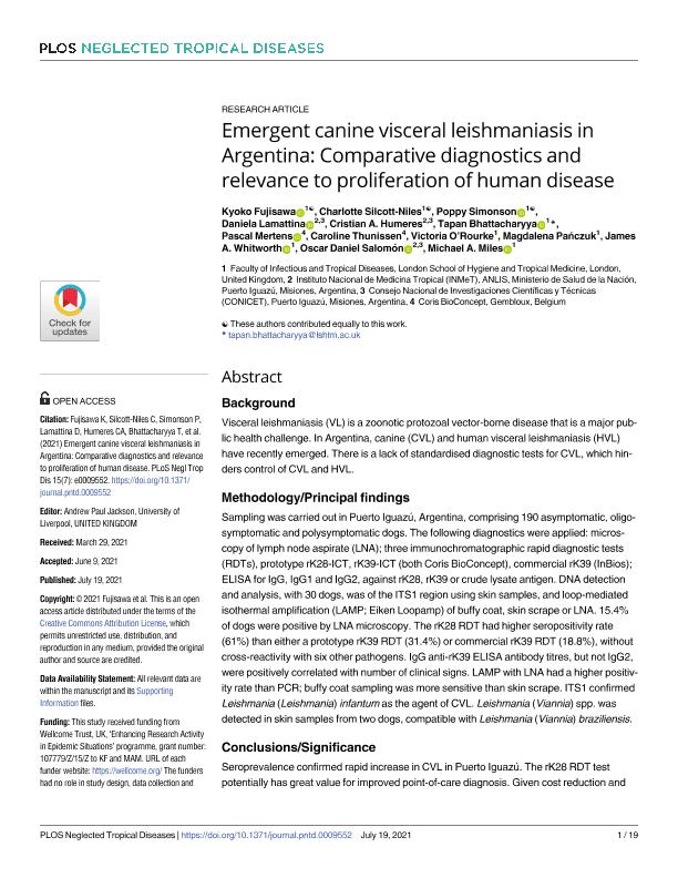
Background Visceral leishmaniasis (VL) is a zoonotic protozoal vector-borne disease that is a major public health challenge. In Argentina, canine (CVL) and human visceral leishmaniasis (HVL) have recently emerged. There is a lack of standardised diagnostic tests for CVL, which hin-ders control of CVL and HVL. Methodology/Principal findings Sampling was carried out in Puerto Iguazú, Argentina, comprising 190 asymptomatic, oligo-symptomatic and polysymptomatic dogs. The following diagnostics were applied: microscopy of lymph node aspirate (LNA); three immunochromatographic rapid diagnostic tests (RDTs), prototype rK28-ICT, rK39-ICT (both Coris BioConcept), commercial rK39 (InBios); ELISA for IgG, IgG1 and IgG2, against rK28, rK39 or crude lysate antigen. DNA detection and analysis, with 30 dogs, was of the ITS1 region using skin samples, and loop-mediated isothermal amplification (LAMP; Eiken Loopamp) of buffy coat, skin scrape or LNA. 15.4% of dogs were positive by LNA microscopy. The rK28 RDT had higher seropositivity rate (61%) than either a prototype rK39 RDT (31.4%) or commercial rK39 RDT (18.8%), without cross-reactivity with six other pathogens. IgG anti-rK39 ELISA antibody titres, but not IgG2, were positively correlated with number of clinical signs. LAMP with LNA had a higher positiv-ity rate than PCR; buffy coat sampling was more sensitive than skin scrape. ITS1 confirmed Leishmania (Leishmania) infantum as the agent of CVL. Leishmania (Viannia) spp. was detected in skin samples from two dogs, compatible with Leishmania (Viannia) braziliensis. Conclusions/Significance Seroprevalence confirmed rapid increase in CVL in Puerto Iguazú. The rK28 RDT test potentially has great value for improved point-of-care diagnosis. Given cost reduction and accessibility, commercial LAMP may be applicable to buffy coat. RDT biomarkers of CVL clinical status are required to combat spread of CVL and HVL. The presence of Viannia, per-haps as an agent of human mucocutaneous leishmaniasis (MCL), highlights the need for vigilance and surveillance.
dc.format
application/pdf
dc.language.iso
eng
dc.publisher
Public Library of Science

dc.rights
info:eu-repo/semantics/openAccess
dc.rights.uri
https://creativecommons.org/licenses/by-nc-sa/2.5/ar/
dc.subject
CANINE VISCERAL LEISHMANIASIS
dc.subject
DIAGNOSIS
dc.subject
LAMP
dc.subject
ARGENTINA
dc.subject.classification
Medicina Tropical

dc.subject.classification
Ciencias de la Salud

dc.subject.classification
CIENCIAS MÉDICAS Y DE LA SALUD

dc.title
Emergent canine visceral leishmaniasis in Argentina: Comparative diagnostics and relevance to proliferation of human disease
dc.type
info:eu-repo/semantics/article
dc.type
info:ar-repo/semantics/artículo
dc.type
info:eu-repo/semantics/publishedVersion
dc.date.updated
2023-08-11T10:44:18Z
dc.journal.volume
15
dc.journal.number
7
dc.journal.pagination
1-19
dc.journal.pais
Estados Unidos

dc.journal.ciudad
San Francisco
dc.description.fil
Fil: Fujisawa, Kyoko. London School of Hygiene and Tropical Medicine; Reino Unido
dc.description.fil
Fil: Silcott Niles, Charlotte. London School of Hygiene and Tropical Medicine; Reino Unido
dc.description.fil
Fil: Simonson, Poppy. London School of Hygiene and Tropical Medicine; Reino Unido
dc.description.fil
Fil: Lamattina, Daniela. Administración Nacional de Laboratorios e Institutos de Salud "Dr. Carlos G. Malbrán". Instituto Nacional de Medicina Tropical; Argentina. Consejo Nacional de Investigaciones Científicas y Técnicas. Centro Científico Tecnológico Conicet - Nordeste; Argentina
dc.description.fil
Fil: Humeres, Cristian Alejandro. Administración Nacional de Laboratorios e Institutos de Salud "Dr. Carlos G. Malbrán". Instituto Nacional de Medicina Tropical; Argentina
dc.description.fil
Fil: Bhattacharyya, Tapan. London School of Hygiene and Tropical Medicine; Reino Unido
dc.description.fil
Fil: Mertens, Pascal. No especifíca;
dc.description.fil
Fil: Thunissen, Caroline. No especifíca;
dc.description.fil
Fil: O'Rourke, Victoria. London School of Hygiene and Tropical Medicine; Reino Unido
dc.description.fil
Fil: Panczuk, Magdalena. London School of Hygiene and Tropical Medicine; Reino Unido
dc.description.fil
Fil: Whitworth, James A.. London School of Hygiene and Tropical Medicine; Reino Unido
dc.description.fil
Fil: Salomón, Oscar Daniel. Consejo Nacional de Investigaciones Científicas y Técnicas; Argentina. Administración Nacional de Laboratorios e Institutos de Salud "Dr. Carlos G. Malbrán". Instituto Nacional de Medicina Tropical; Argentina
dc.description.fil
Fil: Miles, Michael A.. London School of Hygiene and Tropical Medicine; Reino Unido
dc.journal.title
Neglected Tropical Diseases

dc.relation.alternativeid
info:eu-repo/semantics/altIdentifier/url/https://dx.plos.org/10.1371/journal.pntd.0009552
dc.relation.alternativeid
info:eu-repo/semantics/altIdentifier/doi/http://dx.doi.org/10.1371/journal.pntd.0009552
Archivos asociados
