Mostrar el registro sencillo del ítem
dc.contributor.author
Ruvinsky, Igor
dc.contributor.author
Katz, Maximiliano Javier

dc.contributor.author
Dreazen, Avigail
dc.contributor.author
Gielchinsky, Yuval
dc.contributor.author
Saada, Ann
dc.contributor.author
Freedman, Nanette
dc.contributor.author
Mishani, Eyal
dc.contributor.author
Zimmerman, Gabriel
dc.contributor.author
Kasir, Judith
dc.contributor.author
Meyuhas, Oded
dc.date.available
2017-07-17T20:57:15Z
dc.date.issued
2009-05
dc.identifier.citation
Ruvinsky, Igor; Katz, Maximiliano Javier; Dreazen, Avigail; Gielchinsky, Yuval; Saada, Ann; et al.; Mice deficient in ribosomal protein S6 phosphorylation suffer from muscle weakness that reflects a growth defect and energy deficit; Public Library of Science; Plos One; 4; 5; 5-2009; e5618
dc.identifier.issn
1932-6203
dc.identifier.uri
http://hdl.handle.net/11336/20753
dc.description.abstract
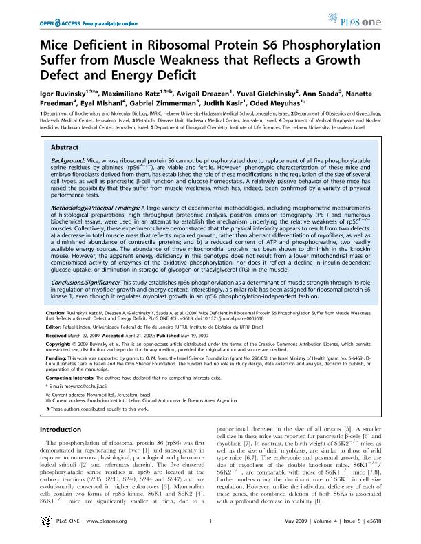
BACKGROUND: Mice, whose ribosomal protein S6 cannot be phosphorylated due to replacement of all five phosphorylatable serine residues by alanines (rpS6(P-/-)), are viable and fertile. However, phenotypic characterization of these mice and embryo fibroblasts derived from them, has established the role of these modifications in the regulation of the size of several cell types, as well as pancreatic beta-cell function and glucose homeostasis. A relatively passive behavior of these mice has raised the possibility that they suffer from muscle weakness, which has, indeed, been confirmed by a variety of physical performance tests. METHODOLOGY/PRINCIPAL FINDINGS: A large variety of experimental methodologies, including morphometric measurements of histological preparations, high throughput proteomic analysis, positron emission tomography (PET) and numerous biochemical assays, were used in an attempt to establish the mechanism underlying the relative weakness of rpS6(P-/-) muscles. Collectively, these experiments have demonstrated that the physical inferiority appears to result from two defects: a) a decrease in total muscle mass that reflects impaired growth, rather than aberrant differentiation of myofibers, as well as a diminished abundance of contractile proteins; and b) a reduced content of ATP and phosphocreatine, two readily available energy sources. The abundance of three mitochondrial proteins has been shown to diminish in the knockin mouse. However, the apparent energy deficiency in this genotype does not result from a lower mitochondrial mass or compromised activity of enzymes of the oxidative phosphorylation, nor does it reflect a decline in insulin-dependent glucose uptake, or diminution in storage of glycogen or triacylglycerol (TG) in the muscle. CONCLUSIONS/SIGNIFICANCE: This study establishes rpS6 phosphorylation as a determinant of muscle strength through its role in regulation of myofiber growth and energy content. Interestingly, a similar role has been assigned for ribosomal protein S6 kinase 1, even though it regulates myoblast growth in an rpS6 phosphorylation-independent fashion.
dc.format
application/pdf
dc.language.iso
eng
dc.publisher
Public Library of Science

dc.rights
info:eu-repo/semantics/openAccess
dc.rights.uri
https://creativecommons.org/licenses/by/2.5/ar/
dc.subject
Muscle Weakness
dc.subject
Glucose Homeostasis
dc.subject
Ribosomal Protein S6
dc.subject
Myoblast
dc.subject.classification
Bioquímica y Biología Molecular

dc.subject.classification
Ciencias Biológicas

dc.subject.classification
CIENCIAS NATURALES Y EXACTAS

dc.title
Mice deficient in ribosomal protein S6 phosphorylation suffer from muscle weakness that reflects a growth defect and energy deficit
dc.type
info:eu-repo/semantics/article
dc.type
info:ar-repo/semantics/artículo
dc.type
info:eu-repo/semantics/publishedVersion
dc.date.updated
2017-07-11T19:28:46Z
dc.identifier.eissn
1932-6203
dc.journal.volume
4
dc.journal.number
5
dc.journal.pagination
e5618
dc.journal.pais
Estados Unidos

dc.journal.ciudad
San Francisco
dc.description.fil
Fil: Ruvinsky, Igor. The Hebrew University Of Jerusalem; Israel
dc.description.fil
Fil: Katz, Maximiliano Javier. The Hebrew University Of Jerusalem; Israel. Consejo Nacional de Investigaciones Científicas y Técnicas. Oficina de Coordinación Administrativa Parque Centenario. Instituto de Investigaciones Bioquímicas de Buenos Aires. Fundación Instituto Leloir. Instituto de Investigaciones Bioquímicas de Buenos Aires; Argentina
dc.description.fil
Fil: Dreazen, Avigail. The Hebrew University Of Jerusalem; Israel
dc.description.fil
Fil: Gielchinsky, Yuval. Hadassah Medical Center; Israel
dc.description.fil
Fil: Saada, Ann. Hadassah Medical Center; Israel
dc.description.fil
Fil: Freedman, Nanette. Hadassah Medical Center; Israel
dc.description.fil
Fil: Mishani, Eyal. Hadassah Medical Center; Israel
dc.description.fil
Fil: Zimmerman, Gabriel. The Hebrew University Of Jerusalem; Israel
dc.description.fil
Fil: Kasir, Judith. The Hebrew University Of Jerusalem; Israel
dc.description.fil
Fil: Meyuhas, Oded. The Hebrew University Of Jerusalem; Israel
dc.journal.title
Plos One

dc.relation.alternativeid
info:eu-repo/semantics/altIdentifier/url/http://journals.plos.org/plosone/article?id=10.1371/journal.pone.0005618
dc.relation.alternativeid
info:eu-repo/semantics/altIdentifier/doi/https://doi.org/10.1371/journal.pone.0005618
Archivos asociados
