Mostrar el registro sencillo del ítem
dc.contributor.author
Vanrell, Maria Cristina

dc.contributor.author
Martinez, Santiago Jose

dc.contributor.author
Muñoz, Lucila Ibel

dc.contributor.author
Salassa, Betiana Nebaí

dc.contributor.author
Gambarte Tudela, Julian Alberto

dc.contributor.author
Romano, Patricia Silvia

dc.date.available
2023-07-26T19:14:50Z
dc.date.issued
2022-07
dc.identifier.citation
Vanrell, Maria Cristina; Martinez, Santiago Jose; Muñoz, Lucila Ibel; Salassa, Betiana Nebaí; Gambarte Tudela, Julian Alberto; et al.; Induction of Autophagy by Ursolic Acid Promotes the Elimination of Trypanosoma cruzi Amastigotes From Macrophages and Cardiac Cells; Frontiers Media; Frontiers in Cellular and Infection Microbiology; 12; 7-2022; 1-12
dc.identifier.issn
2235-2988
dc.identifier.uri
http://hdl.handle.net/11336/205717
dc.description.abstract
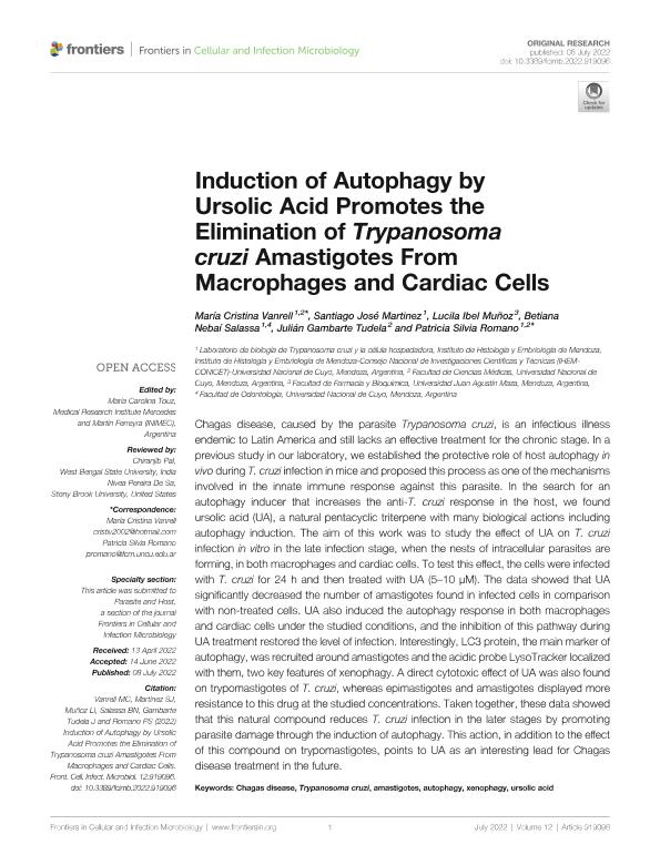
Chagas disease, caused by the parasite Trypanosoma cruzi, is an infectious illness endemic to Latin America and still lacks an effective treatment for the chronic stage. In a previous study in our laboratory, we established the protective role of host autophagy in vivo during T. cruzi infection in mice and proposed this process as one of the mechanisms involved in the innate immune response against this parasite. In the search for an autophagy inducer that increases the anti-T. cruzi response in the host, we found ursolic acid (UA), a natural pentacyclic triterpene with many biological actions including autophagy induction. The aim of this work was to study the effect of UA on T. cruzi infection in vitro in the late infection stage, when the nests of intracellular parasites are forming, in both macrophages and cardiac cells. To test this effect, the cells were infected with T. cruzi for 24 h and then treated with UA (5–10 µM). The data showed that UA significantly decreased the number of amastigotes found in infected cells in comparison with non-treated cells. UA also induced the autophagy response in both macrophages and cardiac cells under the studied conditions, and the inhibition of this pathway during UA treatment restored the level of infection. Interestingly, LC3 protein, the main marker of autophagy, was recruited around amastigotes and the acidic probe LysoTracker localized with them, two key features of xenophagy. A direct cytotoxic effect of UA was also found on trypomastigotes of T. cruzi, whereas epimastigotes and amastigotes displayed more resistance to this drug at the studied concentrations. Taken together, these data showed that this natural compound reduces T. cruzi infection in the later stages by promoting parasite damage through the induction of autophagy. This action, in addition to the effect of this compound on trypomastigotes, points to UA as an interesting lead for Chagas disease treatment in the future.
dc.format
application/pdf
dc.language.iso
eng
dc.publisher
Frontiers Media

dc.rights
info:eu-repo/semantics/openAccess
dc.rights.uri
https://creativecommons.org/licenses/by/2.5/ar/
dc.subject
AMASTIGOTES
dc.subject
AUTOPHAGY
dc.subject
CHAGAS DISEASE
dc.subject
TRYPANOSOMA CRUZI
dc.subject
URSOLIC ACID
dc.subject
XENOPHAGY
dc.subject.classification
Enfermedades Infecciosas

dc.subject.classification
Ciencias de la Salud

dc.subject.classification
CIENCIAS MÉDICAS Y DE LA SALUD

dc.title
Induction of Autophagy by Ursolic Acid Promotes the Elimination of Trypanosoma cruzi Amastigotes From Macrophages and Cardiac Cells
dc.type
info:eu-repo/semantics/article
dc.type
info:ar-repo/semantics/artículo
dc.type
info:eu-repo/semantics/publishedVersion
dc.date.updated
2023-07-18T14:19:44Z
dc.journal.volume
12
dc.journal.pagination
1-12
dc.journal.pais
Suiza

dc.description.fil
Fil: Vanrell, Maria Cristina. Consejo Nacional de Investigaciones Científicas y Técnicas. Centro Científico Tecnológico Conicet - Mendoza. Instituto de Histología y Embriología de Mendoza Dr. Mario H. Burgos. Universidad Nacional de Cuyo. Facultad de Ciencias Médicas. Instituto de Histología y Embriología de Mendoza Dr. Mario H. Burgos; Argentina. Universidad Nacional de Cuyo. Facultad de Ciencias Médicas; Argentina
dc.description.fil
Fil: Martinez, Santiago Jose. Consejo Nacional de Investigaciones Científicas y Técnicas. Centro Científico Tecnológico Conicet - Mendoza. Instituto de Histología y Embriología de Mendoza Dr. Mario H. Burgos. Universidad Nacional de Cuyo. Facultad de Ciencias Médicas. Instituto de Histología y Embriología de Mendoza Dr. Mario H. Burgos; Argentina
dc.description.fil
Fil: Muñoz, Lucila Ibel. Universidad "Juan Agustín Maza"; Argentina
dc.description.fil
Fil: Salassa, Betiana Nebaí. Consejo Nacional de Investigaciones Científicas y Técnicas. Centro Científico Tecnológico Conicet - Mendoza. Instituto de Histología y Embriología de Mendoza Dr. Mario H. Burgos. Universidad Nacional de Cuyo. Facultad de Ciencias Médicas. Instituto de Histología y Embriología de Mendoza Dr. Mario H. Burgos; Argentina. Universidad Nacional de Cuyo. Facultad de Odontologia; Argentina
dc.description.fil
Fil: Gambarte Tudela, Julian Alberto. Consejo Nacional de Investigaciones Científicas y Técnicas. Centro Científico Tecnológico Conicet - Mendoza. Instituto de Medicina y Biología Experimental de Cuyo; Argentina. Universidad Nacional de Cuyo. Facultad de Ciencias Médicas; Argentina
dc.description.fil
Fil: Romano, Patricia Silvia. Consejo Nacional de Investigaciones Científicas y Técnicas. Centro Científico Tecnológico Conicet - Mendoza. Instituto de Histología y Embriología de Mendoza Dr. Mario H. Burgos. Universidad Nacional de Cuyo. Facultad de Ciencias Médicas. Instituto de Histología y Embriología de Mendoza Dr. Mario H. Burgos; Argentina. Universidad Nacional de Cuyo. Facultad de Ciencias Médicas; Argentina
dc.journal.title
Frontiers in Cellular and Infection Microbiology
dc.relation.alternativeid
info:eu-repo/semantics/altIdentifier/url/https://www.frontiersin.org/articles/10.3389/fcimb.2022.919096/full
dc.relation.alternativeid
info:eu-repo/semantics/altIdentifier/doi/http://dx.doi.org/10.3389/fcimb.2022.919096
Archivos asociados
