Artículo
How visual space maps in the optic neuropils of a crab
Fecha de publicación:
04/2011
Editorial:
Wiley-liss, Inc
Revista:
Journal Of Comparative Neurology
ISSN:
0021-9967
Idioma:
Inglés
Tipo de recurso:
Artículo publicado
Clasificación temática:
Resumen
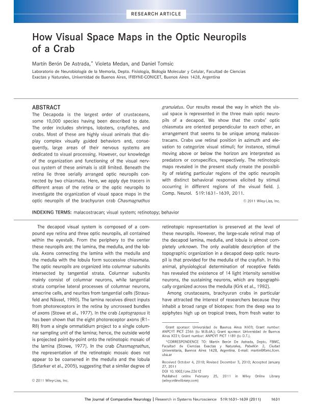
The Decapoda is the largest order of crustaceans, some 10,000 species having been described to date. The order includes shrimps, lobsters, crayfishes, and crabs. Most of these are highly visual animals that display complex visually guided behaviors and, consequently, large areas of their nervous systems are dedicated to visual processing. However, our knowledge of the organization and functioning of the visual nervous system of these animals is still limited. Beneath the retina lie three serially arranged optic neuropils connected by two chiasmata. Here, we apply dye tracers in different areas of the retina or the optic neuropils to investigate the organization of visual space maps in the optic neuropils of the brachyuran crab Chasmagnathus granulatus. Our results reveal the way in which the visual space is represented in the three main optic neuropils of a decapod. We show that the crabs' optic chiasmata are oriented perpendicular to each other, an arrangement that seems to be unique among malacostracans. Crabs use retinal position in azimuth and elevation to categorize visual stimuli; for instance, stimuli moving above or below the horizon are interpreted as predators or conspecifics, respectively. The retinotopic maps revealed in the present study create the possibility of relating particular regions of the optic neuropils with distinct behavioral responses elicited by stimuli occurring in different regions of the visual field.
Palabras clave:
Crustáceos
,
Vision
,
Sistema Nerivioso
,
Retinotopia
Archivos asociados
Licencia
Identificadores
Colecciones
Articulos(IFIBYNE)
Articulos de INST.DE FISIOL., BIOL.MOLECULAR Y NEUROCIENCIAS
Articulos de INST.DE FISIOL., BIOL.MOLECULAR Y NEUROCIENCIAS
Citación
Berón de Astrada, Martín; Medan, Violeta; Tomsic, Daniel; How visual space maps in the optic neuropils of a crab
; Wiley-liss, Inc; Journal Of Comparative Neurology; 519; 9; 4-2011; 1631-1639
Compartir
Altmétricas


