Mostrar el registro sencillo del ítem
dc.contributor.author
Croxatto, Juan Oscar

dc.contributor.author
Tytiun, Adriana E.
dc.contributor.author
Argento, Carlos J.
dc.date.available
2023-03-21T16:01:13Z
dc.date.issued
2010-09
dc.identifier.citation
Croxatto, Juan Oscar; Tytiun, Adriana E.; Argento, Carlos J.; Sequential in vivo confocal microscopy study of corneal wound healing after cross-linking in patients with keratoconus; Slack Inc; JOURNAL OF REFRACTIVE SURGERY - (Print); 26; 9; 9-2010; 638-645
dc.identifier.issn
1081-597X
dc.identifier.uri
http://hdl.handle.net/11336/191228
dc.description.abstract
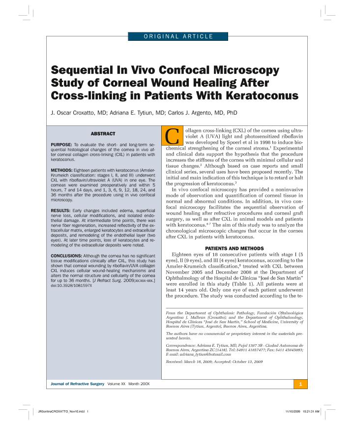
PURPOSE: To evaluate the short- and long-term sequential histological changes of the cornea in vivo after corneal collagen cross-linking (CXL) in patients with keratoconus. METHODS: Eighteen patients with keratoconus (Amsler-Krumeich classification: stages I, II, and III) underwent CXL with riboflavin/ultraviolet A (UVA) in one eye. The corneas were examined preoperatively and within 5 hours, 7 and 14 days, and 1, 3, 6, 9, 12, 18, 24, and 36 months after the procedure using in vivo confocal microscopy. RESULTS: Early changes included edema, superficial nerve loss, cellular modifications, and isolated endothelial damage. At intermediate time points, there was nerve fiber regeneration, increased reflectivity of the extracellular matrix, enlarged keratocytes and extracellular deposits, and remodeling of the endothelial layer (two eyes). At later time points, loss of keratocytes and remodeling of the extracellular deposits were noted. CONCLUSIONS: Although the cornea has no significant tissue modifications clinically after CXL, this study has shown that corneal wounding by riboflavin/UVA collagen CXL induces cellular wound-healing mechanisms and alters the normal structure and cellularity of the cornea for up to 36 months.
dc.format
application/pdf
dc.language.iso
eng
dc.publisher
Slack Inc

dc.rights
info:eu-repo/semantics/openAccess
dc.rights.uri
https://creativecommons.org/licenses/by-nc-sa/2.5/ar/
dc.subject
Crosslinking
dc.subject
Keratoconus
dc.subject
Corneal would healing
dc.subject.classification
Oftalmología

dc.subject.classification
Medicina Clínica

dc.subject.classification
CIENCIAS MÉDICAS Y DE LA SALUD

dc.title
Sequential in vivo confocal microscopy study of corneal wound healing after cross-linking in patients with keratoconus
dc.type
info:eu-repo/semantics/article
dc.type
info:ar-repo/semantics/artículo
dc.type
info:eu-repo/semantics/publishedVersion
dc.date.updated
2023-03-12T15:47:32Z
dc.journal.volume
26
dc.journal.number
9
dc.journal.pagination
638-645
dc.journal.pais
Estados Unidos

dc.description.fil
Fil: Croxatto, Juan Oscar. Fundación Oftalmología Argentina "J. Malbrán"; Argentina. Consejo Nacional de Investigaciones Científicas y Técnicas; Argentina
dc.description.fil
Fil: Tytiun, Adriana E.. Universidad de Buenos Aires; Argentina
dc.description.fil
Fil: Argento, Carlos J.. Universidad de Buenos Aires; Argentina
dc.journal.title
JOURNAL OF REFRACTIVE SURGERY - (Print)

dc.relation.alternativeid
info:eu-repo/semantics/altIdentifier/url/https://journals.healio.com/doi/10.3928/1081597X-20091111-01
dc.relation.alternativeid
info:eu-repo/semantics/altIdentifier/doi/http://dx.doi.org/10.3928/1081597X-20091111-01
Archivos asociados
