Mostrar el registro sencillo del ítem
dc.contributor.author
Castaño, Eduardo Miguel

dc.contributor.author
Maarouf, Chera L.
dc.contributor.author
Wu, Terence
dc.contributor.author
Leal, Maria Celeste

dc.contributor.author
Whiteside, Charisse M.
dc.contributor.author
Lue, Lih-Fen
dc.contributor.author
Kokjohn, Tyler A.
dc.contributor.author
Sabbagh, Marwan N.
dc.contributor.author
Beach, Thomas G.
dc.contributor.author
Roher, Alex E.
dc.date.available
2017-06-16T19:30:50Z
dc.date.issued
2013-01
dc.identifier.citation
Castaño, Eduardo Miguel; Maarouf, Chera L.; Wu, Terence; Leal, Maria Celeste; Whiteside, Charisse M.; et al.; Alzheimer disease periventricular white matter lesions exhibit specific proteomic profile alterations; Elsevier; Neurochemistry International; 62; 2; 1-2013; 145-156
dc.identifier.issn
0197-0186
dc.identifier.uri
http://hdl.handle.net/11336/18335
dc.description.abstract
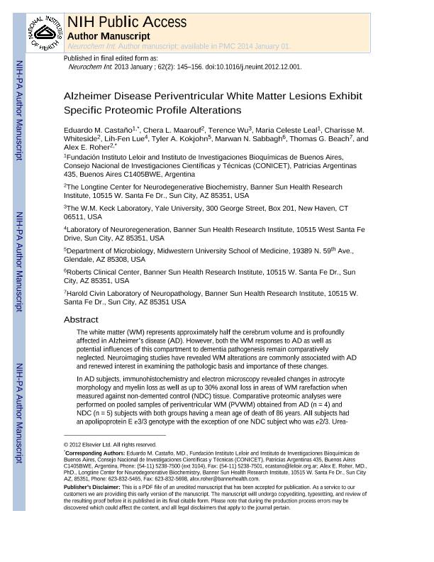
The white matter (WM) represents approximately half the cerebrum volume and is profoundly affected in Alzheimer's disease (AD). However, both the WM responses to AD as well as potential influences of this compartment to dementia pathogenesis remain comparatively neglected. Neuroimaging studies have revealed WM alterations are commonly associated with AD and renewed interest in examining the pathologic basis and importance of these changes. In AD subjects, immunohistochemistry and electron microscopy revealed changes in astrocyte morphology and myelin loss as well as up to 30% axonal loss in areas of WM rarefaction when measured against non-demented control (NDC) tissue. Comparative proteomic analyses were performed on pooled samples of periventricular WM (PVWM) obtained from AD (n=4) and NDC (n=5) subjects with both groups having a mean age of death of 86 years. All subjects had an apolipoprotein E ε3/3 genotype with the exception of one NDC subject who was ε2/3. Urea-detergent homogenates were analyzed using two different separation techniques: 2-dimensional isoelectric focusing/reverse-phase chromatography and 2-dimensional difference gel electrophoresis (2D-DIGE). Proteins with different expression levels between the 2 diagnostic groups were identified using MALDI-Tof/Tof mass spectrometry. In addition, Western blots were used to quantify proteins of interest in individual AD and NDC cases. Our proteomic studies revealed that when WM protein pools were loaded at equal amounts of total protein for comparative analyses, there were quantitative differences between the 2 groups. Molecules related to cytoskeleton maintenance, calcium metabolism and cellular survival such as glial fibrillary acidic protein, vimentin, tropomyosin, collapsin response mediator protein-2, calmodulin, S100-P, annexin A1, α-internexin, α- and β-synuclein, α-B-crystalline, fascin-1, ubiquitin carboxyl-terminal esterase and thymosine were altered between AD and NDC pools. Our experiments suggest that WM activities become globally impaired during the course of AD with significant morphological, biochemical and functional consequential implications for gray matter function and cognitive deficits. These observations may endorse the hypothesis that WM dysfunction is not only a consequence of AD pathology, but that it may precipitate and/or potentiate AD dementia.
dc.format
application/pdf
dc.language.iso
eng
dc.publisher
Elsevier

dc.rights
info:eu-repo/semantics/openAccess
dc.rights.uri
https://creativecommons.org/licenses/by-nc-sa/2.5/ar/
dc.subject
Alzheimer Disease
dc.subject
Periventricular White Matter
dc.subject
White Matter Rarefaction
dc.subject
Proteomics
dc.subject.classification
Neurociencias

dc.subject.classification
Medicina Básica

dc.subject.classification
CIENCIAS MÉDICAS Y DE LA SALUD

dc.title
Alzheimer disease periventricular white matter lesions exhibit specific proteomic profile alterations
dc.type
info:eu-repo/semantics/article
dc.type
info:ar-repo/semantics/artículo
dc.type
info:eu-repo/semantics/publishedVersion
dc.date.updated
2016-09-05T13:17:51Z
dc.identifier.eissn
1872-9754
dc.journal.volume
62
dc.journal.number
2
dc.journal.pagination
145-156
dc.journal.pais
Reino Unido

dc.journal.ciudad
Oxford
dc.description.fil
Fil: Castaño, Eduardo Miguel. Consejo Nacional de Investigaciones Científicas y Técnicas. Oficina de Coordinación Administrativa Parque Centenario. Instituto de Investigaciones Bioquímicas de Buenos Aires. Fundación Instituto Leloir. Instituto de Investigaciones Bioquímicas de Buenos Aires; Argentina
dc.description.fil
Fil: Maarouf, Chera L.. Banner Sun Health Research Institute; Estados Unidos
dc.description.fil
Fil: Wu, Terence. University of Yale; Estados Unidos
dc.description.fil
Fil: Leal, Maria Celeste. Consejo Nacional de Investigaciones Científicas y Técnicas. Oficina de Coordinación Administrativa Parque Centenario. Instituto de Investigaciones Bioquímicas de Buenos Aires. Fundación Instituto Leloir. Instituto de Investigaciones Bioquímicas de Buenos Aires; Argentina
dc.description.fil
Fil: Whiteside, Charisse M.. Banner Sun Health Research Institute; Estados Unidos
dc.description.fil
Fil: Lue, Lih-Fen. Banner Sun Health Research Institute; Estados Unidos
dc.description.fil
Fil: Kokjohn, Tyler A.. Midwestern University School of Medicine; Estados Unidos
dc.description.fil
Fil: Sabbagh, Marwan N.. Banner Sun Health Research Institute; Estados Unidos
dc.description.fil
Fil: Beach, Thomas G.. Banner Sun Health Research Institute; Estados Unidos
dc.description.fil
Fil: Roher, Alex E.. Banner Sun Health Research Institute; Estados Unidos
dc.journal.title
Neurochemistry International

dc.relation.alternativeid
info:eu-repo/semantics/altIdentifier/url/http://www.sciencedirect.com/science/article/pii/S0197018612003919
dc.relation.alternativeid
info:eu-repo/semantics/altIdentifier/doi/https://doi.org/10.1016/j.neuint.2012.12.001
Archivos asociados
