Mostrar el registro sencillo del ítem
dc.contributor.author
Waisman, Ariel

dc.contributor.author
Norris, Alessandra

dc.contributor.author
Elias Costa, Martin

dc.contributor.author
Kopinke, Daniel
dc.date.available
2022-12-16T19:31:44Z
dc.date.issued
2021-12
dc.identifier.citation
Waisman, Ariel; Norris, Alessandra; Elias Costa, Martin; Kopinke, Daniel; Automatic and unbiased segmentation and quantification of myofibers in skeletal muscle; Nature; Scientific Reports; 11; 1; 12-2021; 1-14; 11793
dc.identifier.issn
2045-2322
dc.identifier.uri
http://hdl.handle.net/11336/181627
dc.description.abstract
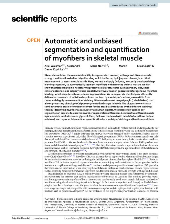
Skeletal muscle has the remarkable ability to regenerate. However, with age and disease muscle strength and function decline. Myofiber size, which is affected by injury and disease, is a critical measurement to assess muscle health. Here, we test and apply Cellpose, a recently developed deep learning algorithm, to automatically segment myofibers within murine skeletal muscle. We first show that tissue fixation is necessary to preserve cellular structures such as primary cilia, small cellular antennae, and adipocyte lipid droplets. However, fixation generates heterogeneous myofiber labeling, which impedes intensity-based segmentation. We demonstrate that Cellpose efficiently delineates thousands of individual myofibers outlined by a variety of markers, even within fixed tissue with highly uneven myofiber staining. We created a novel ImageJ plugin (LabelsToRois) that allows processing of multiple Cellpose segmentation images in batch. The plugin also contains a semi-automatic erosion function to correct for the area bias introduced by the different stainings, thereby identifying myofibers as accurately as human experts. We successfully applied our segmentation pipeline to uncover myofiber regeneration differences between two different muscle injury models, cardiotoxin and glycerol. Thus, Cellpose combined with LabelsToRois allows for fast, unbiased, and reproducible myofiber quantification for a variety of staining and fixation conditions.
dc.format
application/pdf
dc.language.iso
eng
dc.publisher
Nature

dc.rights
info:eu-repo/semantics/openAccess
dc.rights.uri
https://creativecommons.org/licenses/by/2.5/ar/
dc.subject
SKELETAL MUSCLE
dc.subject
IMAGE ANALYSIS
dc.subject
CELL SEGMENTATION
dc.subject.classification
Biología Celular, Microbiología

dc.subject.classification
Ciencias Biológicas

dc.subject.classification
CIENCIAS NATURALES Y EXACTAS

dc.title
Automatic and unbiased segmentation and quantification of myofibers in skeletal muscle
dc.type
info:eu-repo/semantics/article
dc.type
info:ar-repo/semantics/artículo
dc.type
info:eu-repo/semantics/publishedVersion
dc.date.updated
2022-08-02T17:19:56Z
dc.journal.volume
11
dc.journal.number
1
dc.journal.pagination
1-14; 11793
dc.journal.pais
Reino Unido

dc.description.fil
Fil: Waisman, Ariel. Fundacion P/la Lucha C/enferm.neurologicas Infancia. Instituto de Neurociencias. - Consejo Nacional de Investigaciones Cientificas y Tecnicas. Oficina de Coordinacion Administrativa Ciudad Universitaria. Instituto de Neurociencias.; Argentina
dc.description.fil
Fil: Norris, Alessandra. University of Florida; Estados Unidos
dc.description.fil
Fil: Elias Costa, Martin. Universidad de Buenos Aires. Facultad de Ciencias Exactas y Naturales; Argentina. Consejo Nacional de Investigaciones Científicas y Técnicas; Argentina
dc.description.fil
Fil: Kopinke, Daniel. University of Florida; Estados Unidos
dc.journal.title
Scientific Reports
dc.relation.alternativeid
info:eu-repo/semantics/altIdentifier/doi/https://doi.org/10.1038/s41598-021-91191-6
dc.relation.alternativeid
info:eu-repo/semantics/altIdentifier/url/https://www.nature.com/articles/s41598-021-91191-6
Archivos asociados
