Mostrar el registro sencillo del ítem
dc.contributor.author
Conrad, Melanie L.

dc.contributor.author
Freitag, Nancy

dc.contributor.author
Diessler, Mónica Elizabeth

dc.contributor.author
Hernandez, Rocío Belén

dc.contributor.author
Barrientos, Gabriela Laura

dc.contributor.author
Rose, Matthias

dc.contributor.author
Casas, Luciano A.
dc.contributor.author
Barbeito, Claudio Gustavo

dc.contributor.author
Blois, Sandra M.

dc.date.available
2022-12-05T12:43:57Z
dc.date.issued
2015-11
dc.identifier.citation
Conrad, Melanie L.; Freitag, Nancy; Diessler, Mónica Elizabeth; Hernandez, Rocío Belén; Barrientos, Gabriela Laura; et al.; Differential spatiotemporal patterns of galectin expression are a hallmark of endotheliochorial placentation; Wiley Blackwell Publishing, Inc; American Journal of Reproductive Immunology; 75; 3; 11-2015; 317-325
dc.identifier.issn
1046-7408
dc.identifier.uri
http://hdl.handle.net/11336/180123
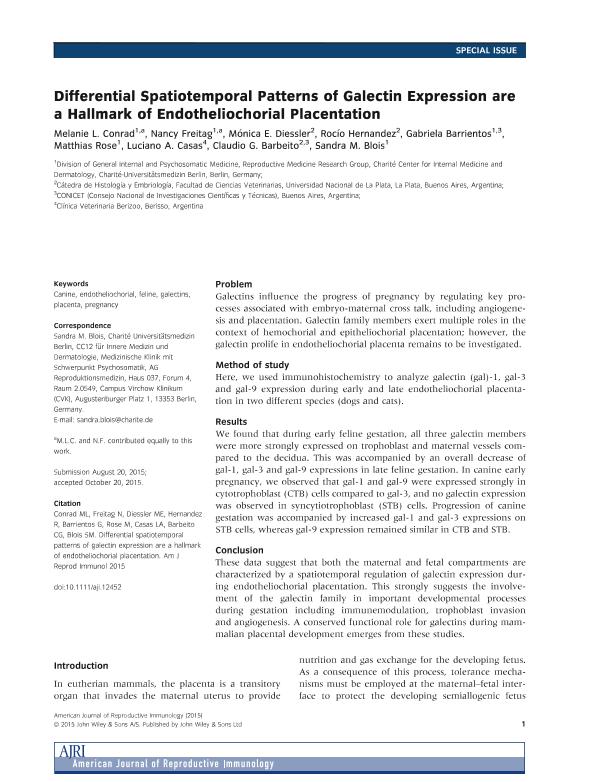
dc.description.abstract
Problem: Galectins influence the progress of pregnancy by regulating key processes associated with embryo-maternal cross talk, including angiogenesis and placentation. Galectin family members exert multiple roles in the context of hemochorial and epitheliochorial placentation; however, the galectin prolife in endotheliochorial placenta remains to be investigated. Method of study: Here, we used immunohistochemistry to analyze galectin (gal)-1, gal-3 and gal-9 expression during early and late endotheliochorial placentation in two different species (dogs and cats). Results: We found that during early feline gestation, all three galectin members were more strongly expressed on trophoblast and maternal vessels compared to the decidua. This was accompanied by an overall decrease of gal-1, gal-3 and gal-9 expressions in late feline gestation. In canine early pregnancy, we observed that gal-1 and gal-9 were expressed strongly in cytotrophoblast (CTB) cells compared to gal-3, and no galectin expression was observed in syncytiotrophoblast (STB) cells. Progression of canine gestation was accompanied by increased gal-1 and gal-3 expressions on STB cells, whereas gal-9 expression remained similar in CTB and STB. Conclusion: These data suggest that both the maternal and fetal compartments are characterized by a spatiotemporal regulation of galectin expression during endotheliochorial placentation. This strongly suggests the involvement of the galectin family in important developmental processes during gestation including immunemodulation, trophoblast invasion and angiogenesis. A conserved functional role for galectins during mammalian placental development emerges from these studies.
dc.format
application/pdf
dc.language.iso
eng
dc.publisher
Wiley Blackwell Publishing, Inc

dc.rights
info:eu-repo/semantics/openAccess
dc.rights.uri
https://creativecommons.org/licenses/by-nc-sa/2.5/ar/
dc.subject
CANINE
dc.subject
ENDOTHELIOCHORIAL
dc.subject
FELINE
dc.subject
GALECTINS
dc.subject
PLACENTA
dc.subject
PREGNANCY
dc.subject.classification
Bioquímica y Biología Molecular

dc.subject.classification
Ciencias Biológicas

dc.subject.classification
CIENCIAS NATURALES Y EXACTAS

dc.subject.classification
Biología del Desarrollo

dc.subject.classification
Ciencias Biológicas

dc.subject.classification
CIENCIAS NATURALES Y EXACTAS

dc.title
Differential spatiotemporal patterns of galectin expression are a hallmark of endotheliochorial placentation
dc.type
info:eu-repo/semantics/article
dc.type
info:ar-repo/semantics/artículo
dc.type
info:eu-repo/semantics/publishedVersion
dc.date.updated
2022-12-05T10:43:33Z
dc.journal.volume
75
dc.journal.number
3
dc.journal.pagination
317-325
dc.journal.pais
Reino Unido

dc.journal.ciudad
Londres
dc.description.fil
Fil: Conrad, Melanie L.. Charité-Universitätsmedizin Berlin; Alemania
dc.description.fil
Fil: Freitag, Nancy. Charité-Universitätsmedizin Berlin; Alemania
dc.description.fil
Fil: Diessler, Mónica Elizabeth. Universidad Nacional de La Plata. Facultad de Ciencias Veterinarias. Departamento de Ciencias Básicas. Cátedra de Histología y Embriología; Argentina
dc.description.fil
Fil: Hernandez, Rocío Belén. Consejo Nacional de Investigaciones Científicas y Técnicas; Argentina. Universidad Nacional de La Plata. Facultad de Ciencias Veterinarias. Departamento de Ciencias Básicas. Cátedra de Histología y Embriología; Argentina
dc.description.fil
Fil: Barrientos, Gabriela Laura. Consejo Nacional de Investigaciones Científicas y Técnicas; Argentina. Charité-Universitätsmedizin Berlin; Alemania
dc.description.fil
Fil: Rose, Matthias. Charité-Universitätsmedizin Berlin; Alemania
dc.description.fil
Fil: Casas, Luciano A.. Clínica Veterinaria Berizoo; Argentina
dc.description.fil
Fil: Barbeito, Claudio Gustavo. Consejo Nacional de Investigaciones Científicas y Técnicas; Argentina. Universidad Nacional de La Plata. Facultad de Ciencias Veterinarias. Departamento de Ciencias Básicas. Cátedra de Histología y Embriología; Argentina
dc.description.fil
Fil: Blois, Sandra M.. Charité-Universitätsmedizin Berlin; Alemania
dc.journal.title
American Journal of Reproductive Immunology

dc.relation.alternativeid
info:eu-repo/semantics/altIdentifier/url/https://onlinelibrary.wiley.com/doi/10.1111/aji.12452
dc.relation.alternativeid
info:eu-repo/semantics/altIdentifier/doi/http://dx.doi.org/10.1111/aji.12452
Archivos asociados
