Mostrar el registro sencillo del ítem
dc.contributor.author
Rodriguez Celin, Alejandra Jimena

dc.contributor.author
Rapacioli, Melina

dc.contributor.author
Gonzalez, Mariela Azul

dc.contributor.author
Ballarin, Virginia Laura

dc.contributor.author
Fiszer, Sara

dc.contributor.author
López, Juan José

dc.contributor.author
Flores, Domingo Vladimir

dc.date.available
2017-06-08T18:48:24Z
dc.date.issued
2015-01
dc.identifier.citation
Rodriguez Celin, Alejandra Jimena; Rapacioli, Melina; Gonzalez, Mariela Azul; Ballarin, Virginia Laura; Fiszer, Sara; et al.; Temporal-spatial correlation between angiogenesis and corticogenesis in the developing chick optic tectum; Public Library of Science; Plos One; 10; 3; 1-2015; 1-24; e0122549
dc.identifier.issn
1932-6203
dc.identifier.uri
http://hdl.handle.net/11336/17793
dc.description.abstract
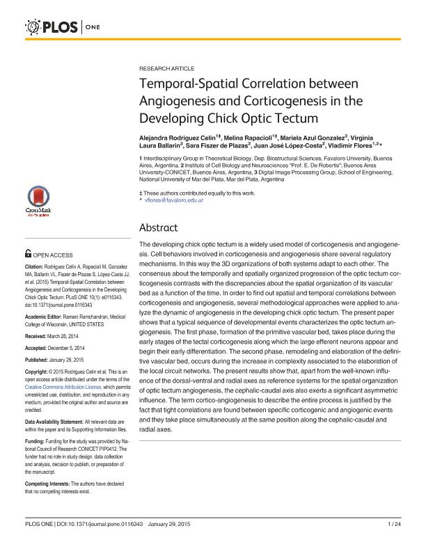
The developing chick optic tectum is a widely used model of corticogenesis and angiogenesis. Cell behaviors involved in corticogenesis and angiogenesis share several regulatory mechanisms. In this way the 3D organizations of both systems adapt to each other. The consensus about the temporally and spatially organized progression of the optic tectum corticogenesis contrasts with the discrepancies about the spatial organization of its vascular bed as a function of the time. In order to find out spatial and temporal correlations between corticogenesis and angiogenesis, several methodological approaches were applied to analyze the dynamic of angiogenesis in the developing chick optic tectum. The present paper shows that a typical sequence of developmental events characterizes the optic tectum angiogenesis. The first phase, formation of the primitive vascular bed, takes place during the early stages of the tectal corticogenesis along which the large efferent neurons appear and begin their early differentiation. The second phase, remodeling and elaboration of the definitive vascular bed, occurs during the increase in complexity associated to the elaboration of the local circuit networks. The present results show that, apart from the well-known influence of the dorsal-ventral and radial axes as reference systems for the spatial organization of optic tectum angiogenesis, the cephalic-caudal axis also exerts a significant asymmetric influence. The term cortico-angiogenesis to describe the entire process is justified by the fact that tight correlations are found between specific corticogenic and angiogenic events and they take place simultaneously at the same position along the cephalic-caudal and radial axes.
dc.format
application/pdf
dc.language.iso
eng
dc.publisher
Public Library of Science

dc.rights
info:eu-repo/semantics/openAccess
dc.rights.uri
https://creativecommons.org/licenses/by/2.5/ar/
dc.subject
Angiogenesis
dc.subject
Neuronal Differentiation
dc.subject
Embryos
dc.subject
Digital Imaging
dc.subject
Superior Colliculus
dc.subject.classification
Ciencias de la Información y Bioinformática

dc.subject.classification
Ciencias de la Computación e Información

dc.subject.classification
CIENCIAS NATURALES Y EXACTAS

dc.subject.classification
Biología del Desarrollo

dc.subject.classification
Ciencias Biológicas

dc.subject.classification
CIENCIAS NATURALES Y EXACTAS

dc.title
Temporal-spatial correlation between angiogenesis and corticogenesis in the developing chick optic tectum
dc.type
info:eu-repo/semantics/article
dc.type
info:ar-repo/semantics/artículo
dc.type
info:eu-repo/semantics/publishedVersion
dc.date.updated
2017-06-07T20:38:13Z
dc.journal.volume
10
dc.journal.number
3
dc.journal.pagination
1-24; e0122549
dc.journal.pais
Estados Unidos

dc.journal.ciudad
San Francisco
dc.description.fil
Fil: Rodriguez Celin, Alejandra Jimena. Universidad Favaloro. Facultad de Medicina. Departamento de Ciencias Bioestructurales. Grupo de Investigación en Biología Teórica; Argentina. Consejo Nacional de Investigaciones Científicas y Técnicas; Argentina
dc.description.fil
Fil: Rapacioli, Melina. Universidad Favaloro. Facultad de Medicina. Departamento de Ciencias Bioestructurales. Grupo de Investigación en Biología Teórica; Argentina. Consejo Nacional de Investigaciones Científicas y Técnicas; Argentina
dc.description.fil
Fil: Gonzalez, Mariela Azul. Universidad Nacional de Mar del Plata. Facultad de Ingeniería; Argentina. Consejo Nacional de Investigaciones Científicas y Técnicas; Argentina
dc.description.fil
Fil: Ballarin, Virginia Laura. Universidad Nacional de Mar del Plata. Facultad de Ingeniería; Argentina
dc.description.fil
Fil: Fiszer, Sara. Consejo Nacional de Investigaciones Científicas y Técnicas. Oficina de Coordinación Administrativa Houssay. Instituto de Biología Celular y Neurociencia "Prof. Eduardo de Robertis". Universidad de Buenos Aires. Facultad de Medicina. Instituto de Biología Celular y Neurociencia; Argentina
dc.description.fil
Fil: López, Juan José. Consejo Nacional de Investigaciones Científicas y Técnicas. Oficina de Coordinación Administrativa Houssay. Instituto de Biología Celular y Neurociencia "Prof. Eduardo de Robertis". Universidad de Buenos Aires. Facultad de Medicina. Instituto de Biología Celular y Neurociencia; Argentina
dc.description.fil
Fil: Flores, Domingo Vladimir. Universidad Favaloro. Facultad de Medicina. Departamento de Ciencias Bioestructurales. Grupo de Investigación en Biología Teórica; Argentina. Consejo Nacional de Investigaciones Científicas y Técnicas. Oficina de Coordinación Administrativa Houssay. Instituto de Biología Celular y Neurociencia "Prof. Eduardo de Robertis". Universidad de Buenos Aires. Facultad de Medicina. Instituto de Biología Celular y Neurociencia; Argentina
dc.journal.title
Plos One

dc.relation.alternativeid
info:eu-repo/semantics/altIdentifier/url/http://journals.plos.org/plosone/article?id=10.1371/journal.pone.0116343
dc.relation.alternativeid
info:eu-repo/semantics/altIdentifier/doi/http://dx.doi.org/10.1371/journal.pone.0116343
Archivos asociados
