Mostrar el registro sencillo del ítem
dc.contributor.author
Luaces, Juan Pablo

dc.contributor.author
Rossi, Luis Francisco

dc.contributor.author
Sciurano, Roberta Beatriz

dc.contributor.author
Rebuzzini, Paola

dc.contributor.author
Merico, Valeria

dc.contributor.author
Zuccotti, Maurizio

dc.contributor.author
Merani, Maria Susana

dc.contributor.author
Garagna, Silvia

dc.date.available
2017-05-26T18:46:29Z
dc.date.issued
2014-01
dc.identifier.citation
Luaces, Juan Pablo; Rossi, Luis Francisco; Sciurano, Roberta Beatriz; Rebuzzini, Paola; Merico, Valeria; et al.; Loss of Sertoli-Germ Cell Adhesion Determines the Rapid Germ Cell Elimination During the Seasonal Regression of the Seminiferous Epithelium of the Long Hairy Armadillo Chaetophractus villosus; Oxford University Press; Biology of Reproduction; 90; 48; 1-2014; 1-11
dc.identifier.issn
0006-3363
dc.identifier.uri
http://hdl.handle.net/11336/16985
dc.description.abstract
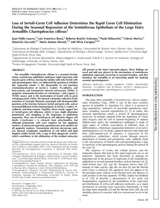
The armadillo Chaetophractus villosus is a seasonal breeder whose seminiferous epithelium undergoes rapid regression with massive germ cell loss, leaving the tubules with only Sertoli cells and spermatogonia. Here, we addressed the question of whether this regression entails 1) the disassembly of cell junctions (immunolocalization of nectin-3, Cadm1, N-cadherin, and beta-catenin, and transmission electron microscopy [TEM]); 2) apoptosis (immunolocalization of cytochrome c and caspase 3; TUNEL assay); and 3) the involvement of Sertoli cells in germ cell phagocytosis (TEM). We showed a dramatic reduction in the extension of vimentin filaments associated with desmosomelike junctions at the interface between Sertoli and germ cells, and an increased diffusion of the immunosignals of nectin-3, Cadm1, N-cadherin, and beta-catenin. Together, these results suggest loss of Sertoli-germ cell adhesion, which in turn might determine postmeiotic cell sloughing at the beginning of epithelium regression. Then, loss of Sertoli-germ cell adhesion triggers cell death. Cytochrome c is released from mitochondria, but although postmeiotic cells were negative for late apoptotic markers, at advanced regression spermatocytes were positive for all apoptotic markers. Transmission electron microscopy analysis showed cytoplasmic engulfment of cell debris and lipid droplets within Sertoli cells, a sign of their phagocytic activity, which contributes to the elimination of the residual meiocytes still present in the latest regression phases. These findings are novel and add new players to the mechanisms of seminiferous epithelium regression occurring in seasonal breeders, and they introduce the armadillo as an interesting model for studying seasonal spermatogenesis.
dc.format
application/pdf
dc.language.iso
eng
dc.publisher
Oxford University Press

dc.rights
info:eu-repo/semantics/openAccess
dc.rights.uri
https://creativecommons.org/licenses/by-nc-sa/2.5/ar/
dc.subject
Adhesion Molecules
dc.subject
Anoikis
dc.subject
Apoptosis
dc.subject
Cadm1
dc.subject
Intermediate Filaments
dc.subject
N-Cadherin
dc.subject
Β-Catenin
dc.subject
Nectin-3
dc.subject
Phagocytosis
dc.subject
Seasonal Reproduction
dc.subject
Spermatogenesis
dc.subject
Testosterone
dc.subject.classification
Biología Reproductiva

dc.subject.classification
Ciencias Biológicas

dc.subject.classification
CIENCIAS NATURALES Y EXACTAS

dc.subject.classification
Biología del Desarrollo

dc.subject.classification
Ciencias Biológicas

dc.subject.classification
CIENCIAS NATURALES Y EXACTAS

dc.title
Loss of Sertoli-Germ Cell Adhesion Determines the Rapid Germ Cell Elimination During the Seasonal Regression of the Seminiferous Epithelium of the Long Hairy Armadillo Chaetophractus villosus
dc.type
info:eu-repo/semantics/article
dc.type
info:ar-repo/semantics/artículo
dc.type
info:eu-repo/semantics/publishedVersion
dc.date.updated
2017-05-15T14:44:55Z
dc.identifier.eissn
1529-7268
dc.journal.volume
90
dc.journal.number
48
dc.journal.pagination
1-11
dc.journal.pais
Reino Unido

dc.journal.ciudad
Oxford
dc.description.fil
Fil: Luaces, Juan Pablo. Universidad de Buenos Aires. Facultad de Medicina. Laboratorio de Biología Cromosómica; Argentina. Consejo Nacional de Investigaciones Científicas y Técnicas; Argentina
dc.description.fil
Fil: Rossi, Luis Francisco. Universidad de Buenos Aires. Facultad de Medicina. Laboratorio de Biología Cromosómica; Argentina. Consejo Nacional de Investigaciones Científicas y Técnicas; Argentina
dc.description.fil
Fil: Sciurano, Roberta Beatriz. Universidad de Buenos Aires. Facultad de Medicina. Laboratorio de Biología Cromosómica; Argentina. Consejo Nacional de Investigaciones Científicas y Técnicas; Argentina
dc.description.fil
Fil: Rebuzzini, Paola. Università degli Studi di Pavia; Italia
dc.description.fil
Fil: Merico, Valeria. Università degli Studi di Pavia; Italia
dc.description.fil
Fil: Zuccotti, Maurizio. Università di Parma; Italia
dc.description.fil
Fil: Merani, Maria Susana. Universidad de Buenos Aires. Facultad de Medicina. Laboratorio de Biología Cromosómica; Argentina. Consejo Nacional de Investigaciones Científicas y Técnicas; Argentina
dc.description.fil
Fil: Garagna, Silvia. Università degli Studi di Pavia; Italia
dc.journal.title
Biology of Reproduction

dc.relation.alternativeid
info:eu-repo/semantics/altIdentifier/url/https://academic.oup.com/biolreprod/article-lookup/doi/10.1095/biolreprod.113.113118
dc.relation.alternativeid
info:eu-repo/semantics/altIdentifier/doi/http://dx.doi.org/10.1095/biolreprod.113.113118
Archivos asociados
