Artículo
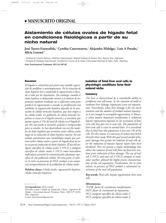
El hígado se caracteriza por poseer una notable capacidad de proliferar y autorregenerarse. En la situación de daño hepático leve o moderado la regeneración es llevada a cabo por los hepatocitos. Sin embargo, cuando el daño hepático es demasiado extenso y el número de hepatocitos maduros residuales no es suficiente como para producir la regeneración o cuando su proliferación está inhibida, la regeneración hepática depende de la activación de células troncales hepáticas, que dan origen a las células ovales. La población de células troncales hepáticas es escasa en el hígado normal y se considera que apenas supera el 1% del total de células en el hígado fetal. Por esta razón es necesario aislarlas y enriquecerlas para su estudio. Se han desarrollado con este fin modelos de daño hepático que permiten aislar células ovales luego de la inducción de daño hepático masivo. En este trabajo presentamos una metodología simple que permite aislar células ovales a partir de hígado fetal de rata sin previa inducción de daño hepático. El uso del antígeno específico de células ovales 2 (OC2) y antígeno específico de células ovales 3 (OC3) como marcadores moleculares permitió la caracterización altamente específica de esta población celular. Por otra parte, el cultivo in vitro en presencia de HGF condujo a un sustancial enriquecimiento de la población de células ovales. The liver is characterized by a remarkable ability to proliferate and self-renew. In the situation of mild or moderate liver damage, hepatocytes carry out regeneration. Nevertheless, when liver damage is far too much extensive and the number of residual mature hepatocytes is not enough to accomplish regeneration, or likewise when mature hepatocyte proliferation is inhibited, hepatic regeneration depends on the activation of liver stem cells that give rise to oval cells. The population of liver stem cells is scant in normal liver. It is considered that in fetal liver this population is just over 1% of the cells. For this reason, it is necessary to isolate and enrich them for their study. With this goal several models of hepatic damage that permit the isolation of oval cells af ter the induction of massive hepatic injure have been developed. Here we present a simple methodology that allows the isolation of oval cells from rat fetal liver without prior induction of liver damage. The use of oval cell 2 (OC2) and oval cell 3 (OC3) antigens as molecular markers allowed the highly precise characterization of this cell population. Furthermore, the in vitro culture in presence of HGF yielded a substantial enrichment of the oval cell population.
Aislamiento de células ovales de hígado fetal en condiciones fisiológicas a partir de su nicho natural
Título:
Isolation of fetal liver oval cells in physiologic conditions form their natural niche
Torres Fuenzalida, Jose Hugo ; Castronuovo, Cynthia Celeste
 ; Hidalgo, Alejandra; Parada, Luis Antonio
 ; Lorenti, Alicia
 ; Castronuovo, Cynthia Celeste
 ; Hidalgo, Alejandra; Parada, Luis Antonio
 ; Lorenti, Alicia
Fecha de publicación:
04/2011
Editorial:
Sociedad Argentina de Gastroenterología
Revista:
Acta Gastroenterologica Latinoamericana.
ISSN:
0300-9033
Idioma:
Español
Tipo de recurso:
Artículo publicado
Clasificación temática:
Resumen
Palabras clave:
Oval Cell
,
Liver
,
Cell Therapy
Archivos asociados
Licencia
Identificadores
Colecciones
Articulos(IPE)
Articulos de INST.DE PATOLOGIA EXPERIMENTAL
Articulos de INST.DE PATOLOGIA EXPERIMENTAL
Citación
Torres Fuenzalida, Jose Hugo; Castronuovo, Cynthia Celeste; Hidalgo, Alejandra; Parada, Luis Antonio; Lorenti, Alicia; Aislamiento de células ovales de hígado fetal en condiciones fisiológicas a partir de su nicho natural; Sociedad Argentina de Gastroenterología; Acta Gastroenterologica Latinoamericana.; 41; 1; 4-2011; 36-46
Compartir


