Mostrar el registro sencillo del ítem
dc.contributor.author
Espinosa, Lady G.
dc.contributor.author
Sosnik, Alejandro Dario

dc.contributor.author
Fontanilla, Marta R.
dc.date.available
2017-04-18T21:57:04Z
dc.date.issued
2010-02
dc.identifier.citation
Espinosa, Lady G.; Sosnik, Alejandro Dario; Fontanilla, Marta R.; Development and preclinical evaluation of acellular collagen scaffolding and autologous artificial connective tissue in the regeneration of oral mucosa wounds; Mary Ann Liebert Inc; Tissue Engineering Part A; 16; 5; 2-2010; 1667-1679
dc.identifier.issn
1937-3341
dc.identifier.uri
http://hdl.handle.net/11336/15426
dc.description.abstract
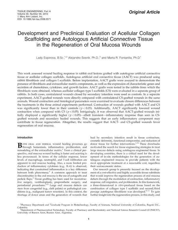
This work assessed wound healing response in rabbit oral lesions grafted with autologous artificial connective tissue or acellular collagen scaffolds. Autologous artificial oral connective tissue (AACT) was produced using rabbit fibroblasts and collagen I scaffolds. Before implantation, AACT grafts were assayed to demonstrate the presence of fibroblasts and extracellular matrix components, as well as the expression of characteristic genes and secretion of chemokines, cytokines, and growth factors. AACT grafts were tested in the rabbits from which the fibroblasts were obtained, whereas acellular collagen type I scaffolds (CS) were evaluated in a separate group of rabbits. In both cases, contralateral wounds closed by secondary intention were used as controls. In a separate experiment, AACT-grafted wounds were directly compared with contralateral CS-grafted wounds in the same animals. Wound contraction and histological parameters were examined to evaluate closure differences between the treatments in the three animal experiments performed. Contraction of wounds grafted with AACT and CS was significantly lower than in their controls (p < 0.05). Additionally, AACT significantly lowered wound contraction when compared with CS (p < 0.05). Intriguingly, it was observed that AACT-grafted wounds initially displayed a significantly higher (p < 0.05)—albeit transient—inflammatory response than seen in CS-grafted wounds and secondary healed wounds. This suggests that an early inflammatory component may contribute to tissue regeneration. Altogether, the results suggest that AACT- and CS-grafted wounds favor regeneration of oral mucosa.
dc.format
application/pdf
dc.language.iso
eng
dc.publisher
Mary Ann Liebert Inc

dc.rights
info:eu-repo/semantics/openAccess
dc.rights.uri
https://creativecommons.org/licenses/by-nc-sa/2.5/ar/
dc.subject
Wound Healing
dc.subject
Animal Models
dc.subject
Dental And Periodontal
dc.subject.classification
Otras Ciencias Químicas

dc.subject.classification
Ciencias Químicas

dc.subject.classification
CIENCIAS NATURALES Y EXACTAS

dc.title
Development and preclinical evaluation of acellular collagen scaffolding and autologous artificial connective tissue in the regeneration of oral mucosa wounds
dc.type
info:eu-repo/semantics/article
dc.type
info:ar-repo/semantics/artículo
dc.type
info:eu-repo/semantics/publishedVersion
dc.date.updated
2017-03-21T20:12:44Z
dc.identifier.eissn
2152-4955
dc.journal.volume
16
dc.journal.number
5
dc.journal.pagination
1667-1679
dc.journal.pais
Estados Unidos

dc.journal.ciudad
Nueva York
dc.description.fil
Fil: Espinosa, Lady G.. Universidad Nacional de Colombia; Colombia
dc.description.fil
Fil: Sosnik, Alejandro Dario. Universidad de Buenos Aires. Facultad de Farmacia y Bioquímica. Departamento de Tecnología Farmacéutica; Argentina. Consejo Nacional de Investigaciones Científicas y Técnicas; Argentina
dc.description.fil
Fil: Fontanilla, Marta R.. Universidad Nacional de Colombia; Colombia
dc.journal.title
Tissue Engineering Part A

dc.relation.alternativeid
info:eu-repo/semantics/altIdentifier/doi/http://dx.doi.org/10.1089/ten.tea.2008.0571
dc.relation.alternativeid
info:eu-repo/semantics/altIdentifier/url/http://online.liebertpub.com/doi/abs/10.1089/ten.tea.2008.0571
Archivos asociados
