Mostrar el registro sencillo del ítem
dc.contributor.author
López Giuliani, Ana C.
dc.contributor.author
Hernández, Eva
dc.contributor.author
Tohmé Chapini, María Julieta

dc.contributor.author
Taisne, Clémence
dc.contributor.author
Roldan, Julieta Suyay

dc.contributor.author
García Samartino, Clara
dc.contributor.author
Lussignol, Marion
dc.contributor.author
Codogno, Patrice
dc.contributor.author
Colombo, María Isabel

dc.contributor.author
Esclatine, Audrey
dc.contributor.author
Delgui, Laura Ruth

dc.date.available
2021-09-30T14:23:02Z
dc.date.issued
2020-09
dc.identifier.citation
López Giuliani, Ana C.; Hernández, Eva; Tohmé Chapini, María Julieta; Taisne, Clémence; Roldan, Julieta Suyay; et al.; Human Cytomegalovirus Inhibits Autophagy of Renal Tubular Epithelial Cells and Promotes Cellular Enlargement; Frontiers Media; Frontiers in Cellular and Infection Microbiology; 10; 9-2020; 1-12
dc.identifier.issn
2235-2988
dc.identifier.uri
http://hdl.handle.net/11336/142069
dc.description.abstract
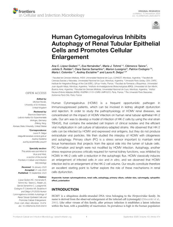
Human Cytomegalovirus (HCMV) is a frequent opportunistic pathogen in immunosuppressed patients, which can be involved in kidney allograft dysfunction and rejection. In order to study the pathophysiology of HCMV renal diseases, we concentrated on the impact of HCMV infection on human renal tubular epithelial HK-2 cells. Our aim was to develop a model of infection of HK-2 cells by using the viral strain TB40/E, that contains the extended cell tropism of clinical isolates and the efficient viral multiplication in cell culture of laboratory-adapted strains. We observed that HK-2 cells can be infected by HCMV and expressed viral antigens, but they do not produce extracellular viral particles. We then studied the interplay of HCMV with ciliogenesis and autophagy. Primary cilium (PC) is a stress sensor important to maintain renal tissue homeostasis that projects from the apical side into the lumen of tubule cells. PC formation and length were not modified by HCMV infection. Autophagy, another stress response process critically required for normal kidney functions, was inhibited by HCMV in HK-2 cells with a reduction in the autophagic flux. HCMV classically induces an enlargement of infected cells in vivo and in vitro, and we observed that HCMV infection led to an enlargement of the HK-2 cell volume. Our results constitute therefore an excellent starting point to further explore the role of these mechanisms in renal cells dysfunction.
dc.format
application/pdf
dc.language.iso
eng
dc.publisher
Frontiers Media

dc.rights
info:eu-repo/semantics/openAccess
dc.rights.uri
https://creativecommons.org/licenses/by/2.5/ar/
dc.subject
AUTOPHAGY
dc.subject
CELLULAR SIZE
dc.subject
CYTOMEGALY
dc.subject
CYTOPATHIC EFFECT
dc.subject
HUMAN CYTOMEGALOVIRUS
dc.subject
POLARIZATION
dc.subject
PRIMARY CILIUM
dc.subject
RENAL CELLS
dc.subject.classification
Biología Celular, Microbiología

dc.subject.classification
Ciencias Biológicas

dc.subject.classification
CIENCIAS NATURALES Y EXACTAS

dc.subject.classification
Virología

dc.subject.classification
Ciencias Biológicas

dc.subject.classification
CIENCIAS NATURALES Y EXACTAS

dc.subject.classification
Enfermedades Infecciosas

dc.subject.classification
Ciencias de la Salud

dc.subject.classification
CIENCIAS MÉDICAS Y DE LA SALUD

dc.title
Human Cytomegalovirus Inhibits Autophagy of Renal Tubular Epithelial Cells and Promotes Cellular Enlargement
dc.type
info:eu-repo/semantics/article
dc.type
info:ar-repo/semantics/artículo
dc.type
info:eu-repo/semantics/publishedVersion
dc.date.updated
2021-09-06T20:02:03Z
dc.journal.volume
10
dc.journal.pagination
1-12
dc.journal.pais
Suiza

dc.description.fil
Fil: López Giuliani, Ana C.. Universidad Nacional de Cuyo. Facultad de Ciencias Exactas y Naturales; Argentina
dc.description.fil
Fil: Hernández, Eva. Centre D'etudes de Saclay; Francia
dc.description.fil
Fil: Tohmé Chapini, María Julieta. Consejo Nacional de Investigaciones Científicas y Técnicas. Centro Científico Tecnológico Conicet - Mendoza. Instituto de Histología y Embriología de Mendoza Dr. Mario H. Burgos. Universidad Nacional de Cuyo. Facultad de Ciencias Médicas. Instituto de Histología y Embriología de Mendoza Dr. Mario H. Burgos; Argentina
dc.description.fil
Fil: Taisne, Clémence. Centre D'etudes de Saclay; Francia
dc.description.fil
Fil: Roldan, Julieta Suyay. Consejo Nacional de Investigaciones Científicas y Técnicas. Centro Científico Tecnológico Conicet - La Plata. Instituto de Investigaciones Biotecnológicas. Universidad Nacional de San Martín. Instituto de Investigaciones Biotecnológicas; Argentina
dc.description.fil
Fil: García Samartino, Clara. Universidad Nacional de Cuyo; Argentina
dc.description.fil
Fil: Lussignol, Marion. Centre D'etudes de Saclay; Francia
dc.description.fil
Fil: Codogno, Patrice. Sorbonne University; Francia
dc.description.fil
Fil: Colombo, María Isabel. Consejo Nacional de Investigaciones Científicas y Técnicas. Centro Científico Tecnológico Conicet - Mendoza. Instituto de Histología y Embriología de Mendoza Dr. Mario H. Burgos. Universidad Nacional de Cuyo. Facultad de Ciencias Médicas. Instituto de Histología y Embriología de Mendoza Dr. Mario H. Burgos; Argentina
dc.description.fil
Fil: Esclatine, Audrey. Centre D'etudes de Saclay; Francia
dc.description.fil
Fil: Delgui, Laura Ruth. Consejo Nacional de Investigaciones Científicas y Técnicas. Centro Científico Tecnológico Conicet - Mendoza. Instituto de Histología y Embriología de Mendoza Dr. Mario H. Burgos. Universidad Nacional de Cuyo. Facultad de Ciencias Médicas. Instituto de Histología y Embriología de Mendoza Dr. Mario H. Burgos; Argentina
dc.journal.title
Frontiers in Cellular and Infection Microbiology
dc.relation.alternativeid
info:eu-repo/semantics/altIdentifier/url/https://www.frontiersin.org/article/10.3389/fcimb.2020.00474/full
dc.relation.alternativeid
info:eu-repo/semantics/altIdentifier/doi/http://dx.doi.org/10.3389/fcimb.2020.00474
Archivos asociados
