Artículo
The Occipital Emissary Vein: A Possible Marker for Pseudotumor Cerebri
Fecha de publicación:
06/2019
Editorial:
American Society of Neuroradiology
Revista:
American Journal Of Neuroradiology
ISSN:
0195-6108
e-ISSN:
1936-959X
Idioma:
Inglés
Tipo de recurso:
Artículo publicado
Clasificación temática:
Resumen
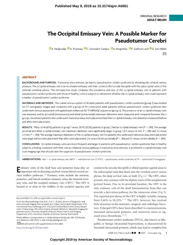
BACKGROUND AND PURPOSE: Transverse sinus stenosis can lead to pseudotumor cerebri syndrome by elevating the cerebral venous pressure. The occipital emissary vein is an inconstant emissary vein that connects the torcular herophili with the suboccipital veins of the external vertebral plexus. This retrospective study compares the prevalence and size of the occipital emissary vein in patients with pseudotumor cerebri syndrome with those in healthy control subjects to determine whether the occipital emissary vein could represent a marker of pseudotumor cerebri syndrome. MATERIALS AND METHODS: The cranial venous system of 46 adult patients with pseudotumor cerebri syndrome (group 1) was studied on CT venography images and compared with a group of 92 consecutive adult patients without pseudotumor cerebri syndrome who underwent venous assessment with gadolinium-enhanced 3D-T1 MPRAGE sequences (group 2). The presence of an occipital emissary vein was assessed, and its proximal (intraosseous) and distal (extracranial) maximum diameters were measured and compared between the 2 groups. Seventeen patients who underwent transverse sinus stent placement had their occipital emissary vein diameters measured before and after stent placement. RESULTS: Thirty of 46 (65%) patients in group 1 versus 29/92 (31.5%) patients in group 2 had an occipital emissary vein (P < .001). The average proximal and distal occipital emissary vein maximum diameters were significantly larger in group 1 (2.3 versus 1.6 mm, P <.005 and 3.3 versus 2.3 mm, P<.001). The average maximum diameters of the occipital emissary vein for patients who underwent transverse sinus stent placement were larger before stent placement than after stent placement: 2.6 versus 1.8 mm proximally (P<.06) and 3.7 versus 2.6 mm distally (P<.005). CONCLUSIONS: Occipital emissary veins are more frequent and larger in patients with pseudotumor cerebri syndrome than in healthy subjects, a finding consistent with their role as collateral venous pathway in transverse sinus stenosis. A prominent occipital emissary vein is an imaging sign that should raise the suspicion of pseudotumor cerebri syndrome.
Palabras clave:
PSEUDOTUMOR CEREBRI
,
EMISSARY VEIN
,
MRI
Archivos asociados
Licencia
Identificadores
Colecciones
Articulos(INCYT)
Articulos de INSTITUTO DE NEUROCIENCIAS COGNITIVAS Y TRASLACIONAL
Articulos de INSTITUTO DE NEUROCIENCIAS COGNITIVAS Y TRASLACIONAL
Citación
Hedjoudje, A.; Piveteau, A.; Gonzalez Campo, Cecilia; Moghekar, A.; Gailloud, P.; et al.; The Occipital Emissary Vein: A Possible Marker for Pseudotumor Cerebri; American Society of Neuroradiology; American Journal Of Neuroradiology; 40; 6; 6-2019; 973-978
Compartir
Altmétricas


