Mostrar el registro sencillo del ítem
dc.contributor.author
Cristofolini, Andrea Lorena

dc.contributor.author
Fiorimanti, Mariana Rita

dc.contributor.author
Campos, M.
dc.contributor.author
Sanchis, Eva Gabriela

dc.contributor.author
Díaz, Tomás

dc.contributor.author
Moschetti, E.
dc.contributor.author
Merkis, Cecilia Inés

dc.date.available
2021-06-07T03:44:26Z
dc.date.issued
2018-02
dc.identifier.citation
Cristofolini, Andrea Lorena; Fiorimanti, Mariana Rita; Campos, M.; Sanchis, Eva Gabriela; Díaz, Tomás; et al.; Morphometric study of the porcine placental vascularization; Wiley Blackwell Publishing, Inc; Reproduction in Domestic Animals; 53; 1; 2-2018; 217-225
dc.identifier.issn
0936-6768
dc.identifier.uri
http://hdl.handle.net/11336/133288
dc.description.abstract
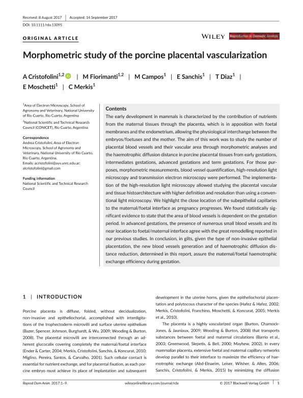
The early development in mammals is characterized by the contribution of nutrients from the maternal tissues through the placenta, which is in apposition with foetal membranes and the endometrium, allowing the physiological interchange between the embryos/foetuses and the mother. The aim of this work was to study the number of placental blood vessels and their vascular area through morphometric analyses and the haemotrophic diffusion distance in porcine placental tissues from early gestations, intermediates gestations, advanced gestations and term gestations. For those purposes, morphometric measurements, blood vessel quantification, high-resolution light microscopy and transmission electron microscopy were performed. The implementation of the high-resolution light microscopy allowed studying the placental vascular and tissue histoarchitecture with higher definition and resolution than using a conventional light microscopy. We highlight the close location of the subepithelial capillaries to the maternal/foetal interface as pregnancy progresses. We found statistically significant evidence to state that the area of blood vessels is dependent on the gestation period. In advanced gestations, the presence of numerous small blood vessels and its near location to foetal/maternal interface agree with the great remodelling reported in our previous studies. In conclusion, in gilts, given the type of non-invasive epithelial placentation, the new blood vessels generation and of haemotrophic diffusion distance reduction, determined in this report, assure the maternal/foetal haemotrophic exchange efficiency during gestation.
dc.format
application/pdf
dc.language.iso
eng
dc.publisher
Wiley Blackwell Publishing, Inc

dc.rights
info:eu-repo/semantics/openAccess
dc.rights.uri
https://creativecommons.org/licenses/by-nc-sa/2.5/ar/
dc.subject
ANGIOGENESIS
dc.subject
GILTS
dc.subject
HAEMOTROPHIC DIFFUSION DISTANCE
dc.subject
VASCULAR AREA
dc.subject.classification
Ciencias Veterinarias

dc.subject.classification
Ciencias Veterinarias

dc.subject.classification
CIENCIAS AGRÍCOLAS

dc.title
Morphometric study of the porcine placental vascularization
dc.type
info:eu-repo/semantics/article
dc.type
info:ar-repo/semantics/artículo
dc.type
info:eu-repo/semantics/publishedVersion
dc.date.updated
2021-06-02T12:14:04Z
dc.identifier.eissn
1439-0531
dc.journal.volume
53
dc.journal.number
1
dc.journal.pagination
217-225
dc.journal.pais
Reino Unido

dc.journal.ciudad
Londres
dc.description.fil
Fil: Cristofolini, Andrea Lorena. Universidad Nacional de Río Cuarto. Facultad de Agronomía y Veterinaria. Departamento de Patología Animal. Área de Microscopia Electrónica; Argentina. Consejo Nacional de Investigaciones Científicas y Técnicas. Centro Científico Tecnológico Conicet - Córdoba; Argentina
dc.description.fil
Fil: Fiorimanti, Mariana Rita. Universidad Nacional de Río Cuarto. Facultad de Agronomía y Veterinaria. Departamento de Patología Animal. Área de Microscopia Electrónica; Argentina. Consejo Nacional de Investigaciones Científicas y Técnicas. Centro Científico Tecnológico Conicet - Córdoba; Argentina
dc.description.fil
Fil: Campos, M.. Universidad Nacional de Río Cuarto. Facultad de Agronomía y Veterinaria. Departamento de Patología Animal. Área de Microscopia Electrónica; Argentina
dc.description.fil
Fil: Sanchis, Eva Gabriela. Universidad Nacional de Río Cuarto. Facultad de Agronomía y Veterinaria. Departamento de Patología Animal. Área de Microscopia Electrónica; Argentina. Consejo Nacional de Investigaciones Científicas y Técnicas. Centro Científico Tecnológico Conicet - Córdoba; Argentina
dc.description.fil
Fil: Díaz, Tomás. Universidad Nacional de Rio Cuarto. Facultad de Agronomia y Veterinaria. Departamento de Anatomia Animal. Laboratorio de Biología Cecular y Embriologia; Argentina. Consejo Nacional de Investigaciones Científicas y Técnicas. Centro Científico Tecnológico Conicet - Córdoba; Argentina
dc.description.fil
Fil: Moschetti, E.. Universidad Nacional de Río Cuarto. Facultad de Agronomía y Veterinaria. Departamento de Patología Animal. Área de Microscopia Electrónica; Argentina
dc.description.fil
Fil: Merkis, Cecilia Inés. Universidad Nacional de Río Cuarto. Facultad de Agronomía y Veterinaria. Departamento de Patología Animal. Área de Microscopia Electrónica; Argentina. Consejo Nacional de Investigaciones Científicas y Técnicas. Centro Científico Tecnológico Conicet - Córdoba; Argentina
dc.journal.title
Reproduction in Domestic Animals

dc.relation.alternativeid
info:eu-repo/semantics/altIdentifier/doi/https://doi.org/10.1111/rda.13095
dc.relation.alternativeid
info:eu-repo/semantics/altIdentifier/url/https://onlinelibrary.wiley.com/doi/full/10.1111/rda.13095
Archivos asociados
