Mostrar el registro sencillo del ítem
dc.contributor.author
Smith, Benjamin R.
dc.contributor.author
Santos, Marta B.
dc.contributor.author
Marshall, Michael S.
dc.contributor.author
Cantuti-Castelvetri, Ludovico
dc.contributor.author
Lopez-Rosas, Aurora
dc.contributor.author
Li, Guannan

dc.contributor.author
Van Breemen, Richard B.

dc.contributor.author
Claycomb, Kumiko I.
dc.contributor.author
Gallea, Jose Ignacio

dc.contributor.author
Celej, Maria Soledad

dc.contributor.author
Crocker, Stephen J.
dc.contributor.author
Givogri, Maria I.
dc.contributor.author
Bongarzone, Ernesto R.
dc.date.available
2021-04-26T21:23:21Z
dc.date.issued
2014-04
dc.identifier.citation
Smith, Benjamin R.; Santos, Marta B.; Marshall, Michael S.; Cantuti-Castelvetri, Ludovico; Lopez-Rosas, Aurora; et al.; Neuronal inclusions of α-synuclein contribute to the pathogenesis of Krabbe disease; John Wiley & Sons Ltd; Journal of Pathology; 232; 5; 4-2014; 509-521
dc.identifier.issn
0022-3417
dc.identifier.uri
http://hdl.handle.net/11336/130864
dc.description.abstract
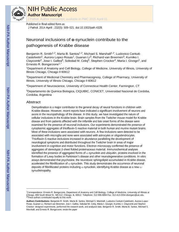
Demyelination is a major contributor to the general decay of neural functions in children with Krabbe disease. However, recent reports have indicated a significant involvement of neurons and axons in the neuropathology of the disease. In this study, we have investigated the nature of cellular inclusions in the Krabbe brain. Brain samples from the twitcher mouse model for Krabbe disease and from patients affected with the infantile and late-onset forms of the disease were examined for the presence of neuronal inclusions. Our experiments demonstrated the presence of cytoplasmic aggregates of thioflavin-S-reactive material in both human and murine mutant brains. Most of these inclusions were associated with neurons. A few inclusions were detected to be associated with microglia and none were associated with astrocytes or oligodendrocytes. Thioflavin-S-reactive inclusions increased in abundance, paralleling the development of neurological symptoms, and distributed throughout the twitcher brain in areas of major involvement in cognition and motor functions. Electron microscopy confirmed the presence of aggregates of stereotypic β-sheet folded proteinaceous material. Immunochemical analyses identified the presence of aggregated forms of α-synuclein and ubiquitin, proteins involved in the formation of Lewy bodies in Parkinson's disease and other neurodegenerative conditions. In vitro assays demonstrated that psychosine, the neurotoxic sphingolipid accumulated in Krabbe disease, accelerated the fibrillization of α-synuclein. This study demonstrates the occurrence of neuronal deposits of fibrillized proteins including α-synuclein, identifying Krabbe disease as a new α-synucleinopathy. Copyright © 2014 Pathological Society of Great Britain and Ireland.
dc.format
application/pdf
dc.language.iso
eng
dc.publisher
John Wiley & Sons Ltd

dc.rights
info:eu-repo/semantics/openAccess
dc.rights.uri
https://creativecommons.org/licenses/by-nc-sa/2.5/ar/
dc.subject
AXONAL DEGENERATION
dc.subject
DYING-BACK PATHOLOGY
dc.subject
KRABBE DISEASE
dc.subject
LEWY BODIES
dc.subject
MYELIN
dc.subject
PSYCHOSINE
dc.subject
SYNUCLEINOPATHIES
dc.subject
UBIQUITIN
dc.subject
Α-SYNUCLEIN
dc.subject.classification
Otras Ciencias Médicas

dc.subject.classification
Otras Ciencias Médicas

dc.subject.classification
CIENCIAS MÉDICAS Y DE LA SALUD

dc.title
Neuronal inclusions of α-synuclein contribute to the pathogenesis of Krabbe disease
dc.type
info:eu-repo/semantics/article
dc.type
info:ar-repo/semantics/artículo
dc.type
info:eu-repo/semantics/publishedVersion
dc.date.updated
2021-03-26T19:37:48Z
dc.identifier.eissn
1096-9896
dc.journal.volume
232
dc.journal.number
5
dc.journal.pagination
509-521
dc.journal.pais
Reino Unido

dc.journal.ciudad
Londres
dc.description.fil
Fil: Smith, Benjamin R.. University Of Ilinois Chicago; Estados Unidos
dc.description.fil
Fil: Santos, Marta B.. University Of Ilinois Chicago; Estados Unidos
dc.description.fil
Fil: Marshall, Michael S.. University Of Ilinois Chicago; Estados Unidos
dc.description.fil
Fil: Cantuti-Castelvetri, Ludovico. University Of Ilinois Chicago; Estados Unidos
dc.description.fil
Fil: Lopez-Rosas, Aurora. University Of Ilinois Chicago; Estados Unidos
dc.description.fil
Fil: Li, Guannan. University Of Ilinois Chicago; Estados Unidos
dc.description.fil
Fil: Van Breemen, Richard B.. University Of Ilinois Chicago; Estados Unidos
dc.description.fil
Fil: Claycomb, Kumiko I.. University of Connecticut; Estados Unidos
dc.description.fil
Fil: Gallea, Jose Ignacio. Consejo Nacional de Investigaciones Científicas y Técnicas. Centro Científico Tecnológico Conicet - Córdoba. Centro de Investigaciones en Química Biológica de Córdoba. Universidad Nacional de Córdoba. Facultad de Ciencias Químicas. Centro de Investigaciones en Química Biológica de Córdoba; Argentina
dc.description.fil
Fil: Celej, Maria Soledad. Consejo Nacional de Investigaciones Científicas y Técnicas. Centro Científico Tecnológico Conicet - Córdoba. Centro de Investigaciones en Química Biológica de Córdoba. Universidad Nacional de Córdoba. Facultad de Ciencias Químicas. Centro de Investigaciones en Química Biológica de Córdoba; Argentina
dc.description.fil
Fil: Crocker, Stephen J.. University of Connecticut; Estados Unidos
dc.description.fil
Fil: Givogri, Maria I.. University of Illinois; Estados Unidos
dc.description.fil
Fil: Bongarzone, Ernesto R.. University of Illinois; Estados Unidos
dc.journal.title
Journal of Pathology

dc.relation.alternativeid
info:eu-repo/semantics/altIdentifier/doi/http://dx.doi.org/10.1002/path.4328
dc.relation.alternativeid
info:eu-repo/semantics/altIdentifier/url/https://onlinelibrary.wiley.com/doi/abs/10.1002/path.4328
dc.relation.alternativeid
info:eu-repo/semantics/altIdentifier/url/https://www.ncbi.nlm.nih.gov/pmc/articles/PMC3977150/
Archivos asociados
