Mostrar el registro sencillo del ítem
dc.contributor.author
Martínez, Gaby F.
dc.contributor.author
Gazal, Nahir Guadalupe

dc.contributor.author
Quassollo Infanzon, Gonzalo Emiliano

dc.contributor.author
Szalai, Alan Marcelo

dc.contributor.author
Del Cid Pellitero, Esther
dc.contributor.author
Durcan, Thomas M.
dc.contributor.author
Fon, Edward A.
dc.contributor.author
Bisbal, Mariano

dc.contributor.author
Stefani, Fernando Daniel

dc.contributor.author
Unsain, Nicolas

dc.date.available
2021-04-15T11:56:21Z
dc.date.issued
2020-02
dc.identifier.citation
Martínez, Gaby F.; Gazal, Nahir Guadalupe; Quassollo Infanzon, Gonzalo Emiliano; Szalai, Alan Marcelo; Del Cid Pellitero, Esther; et al.; Quantitative expansion microscopy for the characterization of the spectrin periodic skeleton of axons using fluorescence microscopy; Nature Publishing Group; Scientific Reports; 10; 1; 2-2020; 1-11
dc.identifier.issn
2045-2322
dc.identifier.uri
http://hdl.handle.net/11336/130089
dc.description.abstract
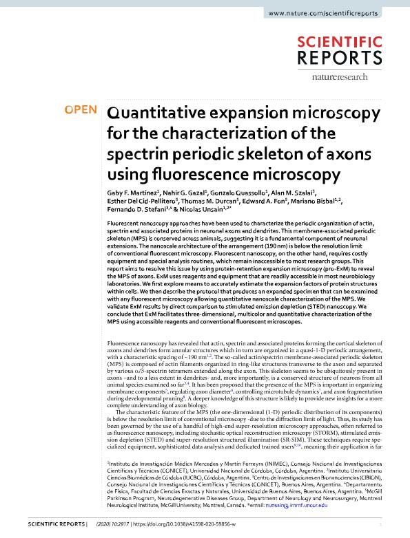
Fluorescent nanoscopy approaches have been used to characterize the periodic organization of actin,spectrin and associated proteins in neuronal axons and dendrites. This membrane-associated periodicskeleton (MPS) is conserved across animals, suggesting it is a fundamental component of neuronalextensions. The nanoscale architecture of the arrangement (190 nm) is below the resolution limitof conventional fluorescent microscopy. Fluorescent nanoscopy, on the other hand, requires costlyequipment and special analysis routines, which remain inaccessible to most research groups. Thisreport aims to resolve this issue by using protein-retention expansion microscopy (pro-ExM) to revealthe MPS of axons. ExM uses reagents and equipment that are readily accessible in most neurobiologylaboratories. We first explore means to accurately estimate the expansion factors of protein structureswithin cells. We then describe the protocol that produces an expanded specimen that can be examinedwith any fluorescent microscopy allowing quantitative nanoscale characterization of the MPS. Wevalidate ExM results by direct comparison to stimulated emission depletion (STED) nanoscopy. Weconclude that ExM facilitates three-dimensional, multicolor and quantitative characterization of theMPS using accessible reagents and conventional fluorescent microscopes.
dc.format
application/pdf
dc.language.iso
eng
dc.publisher
Nature Publishing Group

dc.rights
info:eu-repo/semantics/openAccess
dc.rights.uri
https://creativecommons.org/licenses/by-nc-sa/2.5/ar/
dc.subject
superresolutions
dc.subject
nanoscopy
dc.subject
microscopy
dc.subject
expansion
dc.subject.classification
Biología Celular, Microbiología

dc.subject.classification
Ciencias Biológicas

dc.subject.classification
CIENCIAS NATURALES Y EXACTAS

dc.title
Quantitative expansion microscopy for the characterization of the spectrin periodic skeleton of axons using fluorescence microscopy
dc.type
info:eu-repo/semantics/article
dc.type
info:ar-repo/semantics/artículo
dc.type
info:eu-repo/semantics/publishedVersion
dc.date.updated
2020-10-27T17:47:42Z
dc.identifier.eissn
2045-2322
dc.journal.volume
10
dc.journal.number
1
dc.journal.pagination
1-11
dc.journal.pais
Estados Unidos

dc.description.fil
Fil: Martínez, Gaby F.. Consejo Nacional de Investigaciones Científicas y Técnicas. Centro Científico Tecnológico Conicet - Córdoba. Instituto de Investigación Médica Mercedes y Martín Ferreyra. Universidad Nacional de Córdoba. Instituto de Investigación Médica Mercedes y Martín Ferreyra; Argentina
dc.description.fil
Fil: Gazal, Nahir Guadalupe. Consejo Nacional de Investigaciones Científicas y Técnicas. Centro Científico Tecnológico Conicet - Córdoba. Instituto de Investigación Médica Mercedes y Martín Ferreyra. Universidad Nacional de Córdoba. Instituto de Investigación Médica Mercedes y Martín Ferreyra; Argentina
dc.description.fil
Fil: Quassollo Infanzon, Gonzalo Emiliano. Consejo Nacional de Investigaciones Científicas y Técnicas. Centro Científico Tecnológico Conicet - Córdoba. Instituto de Investigación Médica Mercedes y Martín Ferreyra. Universidad Nacional de Córdoba. Instituto de Investigación Médica Mercedes y Martín Ferreyra; Argentina
dc.description.fil
Fil: Szalai, Alan Marcelo. Consejo Nacional de Investigaciones Científicas y Técnicas. Oficina de Coordinación Administrativa Parque Centenario. Centro de Investigaciones en Bionanociencias "Elizabeth Jares Erijman"; Argentina
dc.description.fil
Fil: Del Cid Pellitero, Esther. No especifíca;
dc.description.fil
Fil: Durcan, Thomas M.. No especifíca;
dc.description.fil
Fil: Fon, Edward A.. No especifíca;
dc.description.fil
Fil: Bisbal, Mariano. Consejo Nacional de Investigaciones Científicas y Técnicas. Centro Científico Tecnológico Conicet - Córdoba. Instituto de Investigación Médica Mercedes y Martín Ferreyra. Universidad Nacional de Córdoba. Instituto de Investigación Médica Mercedes y Martín Ferreyra; Argentina
dc.description.fil
Fil: Stefani, Fernando Daniel. Consejo Nacional de Investigaciones Científicas y Técnicas. Oficina de Coordinación Administrativa Parque Centenario. Centro de Investigaciones en Bionanociencias "Elizabeth Jares Erijman"; Argentina
dc.description.fil
Fil: Unsain, Nicolas. Consejo Nacional de Investigaciones Científicas y Técnicas. Centro Científico Tecnológico Conicet - Córdoba. Instituto de Investigación Médica Mercedes y Martín Ferreyra. Universidad Nacional de Córdoba. Instituto de Investigación Médica Mercedes y Martín Ferreyra; Argentina
dc.journal.title
Scientific Reports
dc.relation.alternativeid
info:eu-repo/semantics/altIdentifier/url/http://www.nature.com/articles/s41598-020-59856-w
dc.relation.alternativeid
info:eu-repo/semantics/altIdentifier/doi/http://dx.doi.org/10.1038/s41598-020-59856-w
Archivos asociados
