Mostrar el registro sencillo del ítem
dc.contributor.author
Porta, Natalia Gabriela

dc.contributor.author
Alvarez, Irene

dc.contributor.author
Suarez Archilla, Guillermo
dc.contributor.author
Ruiz, Vanesa

dc.contributor.author
Abdala, Alejandro
dc.contributor.author
Trono, Karina Gabriela

dc.date.available
2021-01-26T17:48:33Z
dc.date.issued
2019-04
dc.identifier.citation
Porta, Natalia Gabriela; Alvarez, Irene; Suarez Archilla, Guillermo; Ruiz, Vanesa; Abdala, Alejandro; et al.; Experimental infection of sheep with Bovine leukemia virus (BLV): Minimum dose of BLV-FLK cells and cell-free BLV and neutralization activity of natural antibodies; Asociación Argentina de Microbiología; Revista Argentina de Microbiología; 51; 4; 4-2019; 316-323
dc.identifier.issn
0325-7541
dc.identifier.uri
http://hdl.handle.net/11336/123800
dc.description.abstract
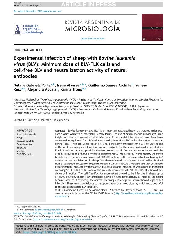
Bovine leukemia virus (BLV) is an important cattle pathogen that causes major economic losses worldwide, especially in dairy farms. The use of animal models provides valuable insight into the pathogenesis of viral infections. Experimental infections of sheep have been conducted using blood from BLV-infected cattle, infectious BLV molecular clones or tumor-derived cells. The Fetal Lamb Kidney cell line, persistently infected with BLV (FLK-BLV), is one of the most commonly used long-term culture available for the permanent production of virus. FLK-BLV cells or the viral particles obtained from the cell-free culture supernatant could be used as a source of provirus or virus to experimentally infect sheep. In this report, we aimed to determine the minimum amount of FLK-BLV cells or cell-free supernatant containing BLV needed to produce infection in sheep. We also evaluated the amount of antibodies obtained from a naturally-infected cow required to neutralize this infection. We observed that both sheep experimentally inoculated with 5000 FLK-BLV cells became infected, as well as one of the sheep receiving 500 FLK-BLV cells. None of the animals inoculated with 50 FLK-BLV cells showed evidence of infection. The cell-free FLK-BLV supernatant proved to be infective in sheep up to a 1:1000 dilution. Specific BLV antibodies showed neutralizing activity as none of the sheep became infected. Conversely, the animals receiving a BLV-negative serum showed signs of BLV infection. These results contribute to the optimization of a sheep bioassay which could be useful to further characterize BLV infection.
dc.description.abstract
El virus de la leucosis bovina (bovine leukemia virus [BLV]) es un importante agente patógeno del ganado que causa importantes pérdidas económicas en todo el mundo, especialmente en los rodeos lecheros. El uso de modelos animales proporciona información valiosa sobre la patogénesis de las infecciones virales. Se realizaron infecciones experimentales en ovejas usando sangre de bovinos infectados con BLV, clones moleculares de BLV infecciosos o células derivadas de tumores. La línea celular Fetal Lamb Kidney, persistentemente infectada con el BLV (FLK-BLV), es uno de los cultivos a largo plazo más utilizados para la producción permanente de virus. Las células FLK-BLV o las partículas virales obtenidas del sobrenadante del cultivo libre de células podrían usarse como fuente de provirus o de virus para infectar experimentalmente ovejas. En este trabajo, nuestro objetivo fue determinar la cantidad mínima de células FLK-BLV o de sobrenadante libre de células que contiene BLV necesaria para producir infección en ovejas. También evaluamos la cantidad de anticuerpos bovinos anti-BLV necesaria para neutralizar la infección. Observamos que las dos ovejas inoculadas experimentalmente con 5000 células FLK-BLV se infectaron, y que una de las dos ovejas que recibieron 500 células FLK-BLV se infectó. Ninguno de los animales inoculados con 50 células FLK-BLV mostró evidencia de infección. El sobrenadante FLK-BLV libre de células demostró ser infectivo en ovejas hasta la dilución 1:1000. Los anticuerpos BLV específicos mostraron actividad neutralizante, ya que ninguna de las ovejas se infectó. Por el contrario, los animales que recibieron un suero BLV negativo mostraron signos de infección por BLV. Estos resultados contribuyen a la optimización de un bioensayo en ovejas útil para caracterizar la infección por BLV.
dc.format
application/pdf
dc.language.iso
eng
dc.publisher
Asociación Argentina de Microbiología

dc.rights
info:eu-repo/semantics/openAccess
dc.rights.uri
https://creativecommons.org/licenses/by-nc-nd/2.5/ar/
dc.subject
Bovine leukemia virus
dc.subject
Animal model
dc.subject
Experimental infection
dc.subject
Sheep
dc.subject
FLK-BLV cells
dc.subject.classification
Otras Ciencias Veterinarias

dc.subject.classification
Ciencias Veterinarias

dc.subject.classification
CIENCIAS AGRÍCOLAS

dc.title
Experimental infection of sheep with Bovine leukemia virus (BLV): Minimum dose of BLV-FLK cells and cell-free BLV and neutralization activity of natural antibodies
dc.title
Infección experimental en ovinos con el virus de leucosis bovina: dosis mínima de células BLV-FLK y virus libre de células, y actividad neutralizante de anticuerpos naturales
dc.type
info:eu-repo/semantics/article
dc.type
info:ar-repo/semantics/artículo
dc.type
info:eu-repo/semantics/publishedVersion
dc.date.updated
2020-11-30T14:17:42Z
dc.identifier.eissn
1851-7617
dc.journal.volume
51
dc.journal.number
4
dc.journal.pagination
316-323
dc.journal.pais
Argentina

dc.description.fil
Fil: Porta, Natalia Gabriela. Instituto Nacional de Tecnología Agropecuaria. Centro de Investigación en Ciencias Veterinarias y Agronómicas. Instituto de Virología; Argentina. Consejo Nacional de Investigaciones Científicas y Técnicas; Argentina
dc.description.fil
Fil: Alvarez, Irene. Instituto Nacional de Tecnología Agropecuaria. Centro de Investigación En Ciencias Veterinarias y Gastronómicas. Instituto de Virología E Innovaciones Tecnológicas. - Consejo Nacional de Investigaciones Científicas y Técnicas. Oficina de Coordinación Administrativa Parque Centenario. Instituto de Virología e Innovaciones Tecnológicas; Argentina
dc.description.fil
Fil: Suarez Archilla, Guillermo. Instituto Nacional de Tecnología Agropecuaria. Centro Regional Santa Fe. Estación Experimental Agropecuaria Rafaela; Argentina
dc.description.fil
Fil: Ruiz, Vanesa. Instituto Nacional de Tecnología Agropecuaria. Centro de Investigación En Ciencias Veterinarias y Gastronómicas. Instituto de Virología E Innovaciones Tecnológicas. - Consejo Nacional de Investigaciones Científicas y Técnicas. Oficina de Coordinación Administrativa Parque Centenario. Instituto de Virología e Innovaciones Tecnológicas; Argentina
dc.description.fil
Fil: Abdala, Alejandro. Instituto Nacional de Tecnología Agropecuaria. Centro Regional Santa Fe. Estación Experimental Agropecuaria Rafaela; Argentina
dc.description.fil
Fil: Trono, Karina Gabriela. Instituto Nacional de Tecnología Agropecuaria. Centro de Investigación En Ciencias Veterinarias y Gastronómicas. Instituto de Virología E Innovaciones Tecnológicas. - Consejo Nacional de Investigaciones Científicas y Técnicas. Oficina de Coordinación Administrativa Parque Centenario. Instituto de Virología e Innovaciones Tecnológicas; Argentina
dc.journal.title
Revista Argentina de Microbiología

dc.relation.alternativeid
info:eu-repo/semantics/altIdentifier/url/https://linkinghub.elsevier.com/retrieve/pii/S0325754119300057
dc.relation.alternativeid
info:eu-repo/semantics/altIdentifier/doi/http://dx.doi.org/10.1016/j.ram.2019.01.004
Archivos asociados
