Mostrar el registro sencillo del ítem
dc.contributor.author
Campoy, Emanuel Martin

dc.contributor.author
Branham, Maria Teresita

dc.contributor.author
Mayorga, Luis Segundo

dc.contributor.author
Roque Moreno, Maria

dc.date.available
2020-12-17T15:49:54Z
dc.date.issued
2019-04
dc.identifier.citation
Campoy, Emanuel Martin; Branham, Maria Teresita; Mayorga, Luis Segundo; Roque Moreno, Maria; Intratumor heterogeneity index of breast carcinomas based on DNA methylation profiles; BioMed Central; BMC Cancer; 19; 1; 4-2019; 1-15
dc.identifier.issn
1471-2407
dc.identifier.uri
http://hdl.handle.net/11336/120774
dc.description.abstract
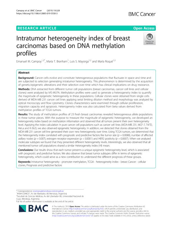
Background: Cancer cells evolve and constitute heterogeneous populations that fluctuate in space and time and are subjected to selection generating intratumor heterogeneity. This phenomenon is determined by the acquisition of genetic/epigenetic alterations and their selection over time which has clinical implications on drug resistance. Methods: DNA extracted from different tumor cell populations (breast carcinomas, cancer cell lines and cellular clones) were analyzed by MS-MLPA. Methylation profiles were used to generate a heterogeneity index to quantify the magnitude of epigenetic heterogeneity in these populations. Cellular clones were obtained from single cells derived of MDA-MB 231 cancer cell lines applying serial limiting dilution method and morphology was analyzed by optical microscopy and flow cytometry. Clones characteristics were examined through cellular proliferation, migration capacity and apoptosis. Heterogeneity index was also calculated from beta values derived from methylation profiles of TCGA tumors. Results: The study of methylation profiles of 23 fresh breast carcinomas revealed heterogeneous allele populations in these tumor pieces. With the purpose to measure the magnitude of epigenetic heterogeneity, we developed an heterogeneity index based on methylation information and observed that all tumors present their own heterogeneity level. Applying the index calculation in pure cancer cell populations such as cancer cell lines (MDA-MB 231, MCF-7, T47D, HeLa and K-562), we also observed epigenetic heterogeneity. In addition, we detected that clones obtained from the MDA-MB 231 cancer cell line generated their own new heterogeneity over time. Using TCGA tumors, we determined that the heterogeneity index correlated with prognostic and predictive factors like tumor size (p = 0.0088), number of affected axillary nodes (p = 0.007), estrogen receptor expression (p < 0.0001) and HER2 positivity (p = 0.0007). When we analyzed molecular subtypes we found that they presented different heterogeneity levels. Interestingly, we also observed that all mentioned tumor cell populations shared a similar Heterogeneity index (HI) mean. Conclusions: Our results show that each tumor presents a unique epigenetic heterogeneity level, which is associated with prognostic and predictive factors. We also observe that breast tumor subtypes differ in terms of epigenetic heterogeneity, which could serve as a new contribution to understand the different prognosis of these groups.
dc.format
application/pdf
dc.language.iso
eng
dc.publisher
BioMed Central

dc.rights
info:eu-repo/semantics/openAccess
dc.rights.uri
https://creativecommons.org/licenses/by-nc-sa/2.5/ar/
dc.subject
INTRATUMOR HETEROGENEITY
dc.subject
PROGNOSIS AND PREDICTIVE FACTORS
dc.subject
TCGA
dc.subject
PROMOTER METHYLATION
dc.subject
HETEROGENEITY INDEX
dc.subject
BREAST CANCER
dc.subject
CELLULAR CLONES
dc.subject.classification
Bioquímica y Biología Molecular

dc.subject.classification
Ciencias Biológicas

dc.subject.classification
CIENCIAS NATURALES Y EXACTAS

dc.title
Intratumor heterogeneity index of breast carcinomas based on DNA methylation profiles
dc.type
info:eu-repo/semantics/article
dc.type
info:ar-repo/semantics/artículo
dc.type
info:eu-repo/semantics/publishedVersion
dc.date.updated
2020-11-19T22:00:24Z
dc.journal.volume
19
dc.journal.number
1
dc.journal.pagination
1-15
dc.journal.pais
Reino Unido

dc.journal.ciudad
Londres
dc.description.fil
Fil: Campoy, Emanuel Martin. Consejo Nacional de Investigaciones Científicas y Técnicas. Centro Científico Tecnológico Conicet - Mendoza. Instituto de Histología y Embriología de Mendoza Dr. Mario H. Burgos. Universidad Nacional de Cuyo. Facultad de Ciencias Médicas. Instituto de Histología y Embriología de Mendoza Dr. Mario H. Burgos; Argentina
dc.description.fil
Fil: Branham, Maria Teresita. Consejo Nacional de Investigaciones Científicas y Técnicas. Centro Científico Tecnológico Conicet - Mendoza. Instituto de Histología y Embriología de Mendoza Dr. Mario H. Burgos. Universidad Nacional de Cuyo. Facultad de Ciencias Médicas. Instituto de Histología y Embriología de Mendoza Dr. Mario H. Burgos; Argentina
dc.description.fil
Fil: Mayorga, Luis Segundo. Consejo Nacional de Investigaciones Científicas y Técnicas. Centro Científico Tecnológico Conicet - Mendoza. Instituto de Histología y Embriología de Mendoza Dr. Mario H. Burgos. Universidad Nacional de Cuyo. Facultad de Ciencias Médicas. Instituto de Histología y Embriología de Mendoza Dr. Mario H. Burgos; Argentina
dc.description.fil
Fil: Roque Moreno, Maria. Consejo Nacional de Investigaciones Científicas y Técnicas. Centro Científico Tecnológico Conicet - Mendoza. Instituto de Histología y Embriología de Mendoza Dr. Mario H. Burgos. Universidad Nacional de Cuyo. Facultad de Ciencias Médicas. Instituto de Histología y Embriología de Mendoza Dr. Mario H. Burgos; Argentina
dc.journal.title
BMC Cancer

dc.relation.alternativeid
info:eu-repo/semantics/altIdentifier/url/https://bmccancer.biomedcentral.com/articles/10.1186/s12885-019-5550-3
dc.relation.alternativeid
info:eu-repo/semantics/altIdentifier/doi/http://dx.doi.org/10.1186/s12885-019-5550-3
Archivos asociados
